Pulmonary Function Testing
Pulmonary Function Testing. CRT 7? = 5% RRT 4?. Which of the following are purposes of assessing pulmonary function? Screen for pulmonary disease Evaluate patients for surgical risk Assess the progression of disease Assist in determining pulmonary disability

Pulmonary Function Testing
E N D
Presentation Transcript
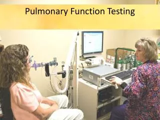
Pulmonary Function Testing CRT 7? = 5% RRT 4?
Which of the following are purposes of assessing pulmonary function? • Screen for pulmonary disease • Evaluate patients for surgical risk • Assess the progression of disease • Assist in determining pulmonary disability • Modify the therapeutic approach to patient care • I, III, and IV • III, IV, and V • I, II, III, IV, and V • II, IV, and V
Which of the following techniques are used to measure RV? • Helium dilution • Body plethysmography • Nitrogen washout • Flow-volume loops • II and IV • I, II, and III • I, II, III, and IV • I, III, and IV
Helium Dilution Closed Method A known % of He is diluted by the patient’s FRC. The change in the He% is used to determine FRC 600 ml 10% He
Nitrogen Washout, Open Method The FRC is washed out of the lung by having the patient inspire 100% O2 to replace the N2 from the FRC. The amount of N2 removed is used to calculate FRC
Boyle’s Law to TGV Patient pants at FRC while pressures and volumes are obtained Raw can be determined by measuring changes in pressure vs. flow 0.6 – 2.4 cmH2O/L/sec Compliance can be determined by measuring the volume change per unit pressure change 60 – 100 mL/cmH2O Plethysmography Body Box
During a helium dilution test for FRC, you notice that it takes 19 minutes for equilibration between the gas concentrations in the spirometer and the patient's lungs. Based on this information, what can you conclude? • The patient has restrictive lung disease. • The spirometer is leaking helium. • The patient has obstructive lung disease. • Insufficient oxygen was added to the system.
What is the gas normally employed to measure the diffusing capacity of the lung? • O2 • CO • CO2 • He
Gas Diffusion (DLCO)Carbon monoxide diffusion capacity • Evaluates diffusion across the A-C membrane • Patient inhales a VC breath of gas containing a known amount of CO. • Breath hold for 10 sec. • Exhaled gas is analyzed. • Normal 25 mLCO/min/mmHg • emphysema, pulmonary fibrosis, sarcoidosis, edema, O2 toxicity
On a patient undergoing testing in the pulmonary function laboratory, you observe a "box–shaped" flow–volume loop with equal reductions in inspiratory and expiratory flows. What does this most likely indicate? • Fixed upper airway obstruction • Variable extrathoracic airway obstruction • Variable intrathoracic airway obstruction • Chronic obstructive pulmonary disease
Severe obstructive disease Flow volume loop from a healthy subject Obstructive airway disease Fixed major airway obstruction Restrictive lung disease
Fixed upper-airway obstruction (intrathoracic or extrathoracic). Variable extrathoracic obstruction. Variable intrathoracic obstruction.
What time period is generally used to measure MVV? • 6 to 8 seconds • 12 to 15 seconds • 30 to 40 seconds • 40 to 60 seconds
Maximum Voluntary Ventilation: tests the ability of the patients chest muscles to expand and contract Pt breaths in and out as fast as possible Normal 170 L/min Decreased in Obstructive dz Increased Raw Muscle weakness Decreased compliance poor patient effort
The best way to check the accuracy of a water-seal spirometer is to use a A.3-L syringe. B.pneumotachometer. C.vortex sensor. D.Wright respirometer.
Calibration • Volume: 3 L syringe • Flow: rotometer • Timing devices: stopwatch • Plethysmograph • Rotometer for flow • Barometer for pressure
After a resting expiration, air still remains in the lungs. What is this volume called? • FRC • VC • RV • ERV
Know your lung volumes and capacities! 3000 ml 3500 ml 4500 ml 1000 ml 2500 ml 1500 ml Memorize numbers from Persing.
During each cycle of normal quiet breathing, a volume of gas is moved into and out of the lungs. What is this cyclical volume called? • IRV • Tidal volume (VT) • ERV • Vital capacity (VC)
Which of the following volumes or capacities cannot be measured by simple spirometry? • Functional residual capacity ( FRC) • Expiratory reserve volume ( ERV) • Residual volume (RV) • Inspiratory reserve volume ( IRV) • I, III, and IV • I, II, III, and IV • I and III • I and IV
Which of the following is equal to total lung capacity (TLC)? • VT + ERV + IRV + RV • IC + VT + ERV • VC + ERV • FRC + IRV
A patient has a VC of 4200 ml, an FRC of 3,300 mL and an ERV of 1500 ml. What is the RV? • 5700 ml • 2700 ml • 1800 ml • 7500 ml
Which of the following is a true statement? • VC = FRC + VT • VC = IRV + VT + ERV • VC = VT + IRV + RV • FRC = VT + ERV
What is the amount of gas that can be inhaled over and above that which is normally inhaled during quiet breathing? • FRC • ERV • IRV • VC
After the most strenuous expiratory effort, air still remains in the lungs and cannot be removed voluntarily. What is this volume called? • IRV • RV • ER • FRC
What is the amount of gas that can be exhaled below the resting expiratory level? • ERV • RV • FRC • VC
Which of the following is the maximum amount of air that can be exhaled from the maximum inspiratory level? A.vital capacity B.residual volume C.functional residual capacity D.expiratory reserve volume
How can you ensure reliability when measuring the ERV? • Have the patient perform the maneuver twice, assure consistency, then take best value. • Have the patient perform the maneuver 3 times, then take the last value. • Have the patient perform the maneuver twice, assure consistency, then take mean value. • Have the patient perform the maneuver until they become fatigued, then take the last value.
A patient has an expired minute ventilation of 14.2 L and a ventilatory rate of 25/min. What is the average VT? • 568 ml • 635 ml • 725 ml • 410 ml
The respiratory therapist instructed a patient to take a deep breath and then exhale as quickly as possible. The therapist observed a recording of the fastest air movement. Which of the following was measured? A. peak flow B. vital capacity C. FEV 1 D. FEF 25-75%
A patient has a prebronchodilator peak expiratory flow rate (PEFR) of 4.5 L/sec. The postbronchodilator value is 5.0 L/sec. What is the percent change? • 11 • 22 • 33 • 50
Peak Flow • Normal • 400 – 600 L/min • 6.5 – 10 L/sec • Percent Change • Post – Pre x 100 Pre • Percent Predicted • Actual x 100 Predicted
Which of the following is being measured if a respiratory care practitioner instructs a patient to take a maximum deep breath and then exhale as much and as fast as possible? • RV • VC • TLC • FVC
FVC = volume Restrictive Obstructive FVC with Normal SVC FEVtime = flow Restrictive Obstructive So look at FEV1/FVC% FEV1/FVC % Normal in Restrictive Obstructive Egan fig. 17-5 97% 75% 60% 94%
A patient has a predicted FEV1 of 4.2 L and a measure FEV1 of 3.5 L. What is the predicted FEV1 in percent? • 76 • 83 • 92 • 120
A patient with chronic obstructive pulmonary disease (COPD) has a normal slow vital capacity (SVC) of 3400 ml and an FVC of 2300 ml. Which of the following mechanisms best explains this difference? • Airway trapping during forced expiration • Muscle fatigue during forced expiration • Decreased compliance during forced expiration • Poor instruction by the pulmonary technologist
Compared to predicted normals, a patient has an increased RV and a decreased percent FEV1/FVC. Test results are repeatable. Which of the following is most likely the underlying problem? • Generalized obstruction with air trapping • Poor patient effort during the test • Restrictive disorder of the lungs • Combined restrictive and obstructive disease
Compared to predicted normals, a patient has a reduced TLC and a decreased percent FEV1/FVC. Test results are repeatable. Which of the following is most likely the underlying problem? • Poor patient effort during the test procedure • Restrictive disorder of the lungs or chest wall • Combined restrictive and obstructive disease • Peripheral (small) airway obstruction
What is the term for the standard measure of the average expiratory flow during the middle portion of an FVC maneuver? • FEV1 • FEF200-1200 • PEFR • FEF25%-75%