Chapter 9 Pulmonary Function Testing
Chapter 9 Pulmonary Function Testing. Overview. PFT includes: Spirometry Flow volume loop (FVL) before and after bronchodilator inhalation Lung volume studies Diffusing capacity (D LCO ) Airway resistance (Raw) Arterial blood gas (ABG) measurements


Chapter 9 Pulmonary Function Testing
E N D
Presentation Transcript
Chapter 9 Pulmonary Function Testing
Overview PFT includes: Spirometry Flow volume loop (FVL) before and after bronchodilator inhalation Lung volume studies Diffusing capacity (DLCO) Airway resistance (Raw) Arterial blood gas (ABG) measurements Pulmonary response to exercise and bronchial provocation
Purpose of PFT Evaluate cause of pulmonary symptoms Evaluate abnormalities seen on the CXR and/or CT scan Follow course of disease and response to treatment Evaluate perioperative risk for pulmonary complications Rule out pulmonary pathology in people with high risk for pulmonary dysfunction Evaluate disability
Normal Values PFT normal values vary with age, height, gender, and race Height the most important factor predicting lung volumes The taller the person, the larger the values Weight important when BMI >30 = restrictive Gender: males have larger lungs Race: African Americans, Asians, East Indians have 12% smaller lung volumes
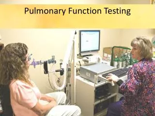
PFT Equipment American Thoracic Society standards Spirometer: routine flows and volume Body plethysmograph: TLC and airway resistance studies Diffusion system: lung diffusion Gas analysis (carbon dioxide, carbon monoxide, helium, nitrogen, and oxygen) Nebulizer equipment for albuterol and methacholine
Arterial blood gas analyzer Treadmill or bicycle for exercise evaluation Laboratories with smaller volumes of tests Multifunction device that measures lung volumes, flow rates, diffusing capacity, and response to bronchial provocation all spirometric values obtained under ambient conditions convert to Body temperature, ambient pressure, saturated (BTPS)
Measures of Lung Function Tidal volume (VT) Residual volume (RV) Expiratory reserve volume (ERV) Inspiratory reserve volume (IRV) Minute volume (VE) Vital capacity (VC) Total lung capacity (TLC) Functional residual capacity (FRC) Inspiratory capacity (IC) Maximal voluntary ventilation (MVV)
Measures of Lung Function (cont’d) Tidal volume Volume during quiet breathing Adults: 350 to 600 ml Stiff lungs: small volumes at higher rate Obstruction: normal volume at slower rate Minute volume Rate x volume 4 to 8 L/min
Measures of Lung Function (cont’d) Vital capacity: maximal volume exhaled Measured after deepest breath possible Slow vital capacity (SVC) Forced vital capacity (FVC) Proper coaching is essential Phases Maximal inspiratory effort Initial expiratory blast Forceful emptying of lungs <20 ml/kg: risk for complications
Measures of Lung Function (cont’d) Total lung capacity Sum of SVC and RV Normal % predicted is 80% to 120% Increased in obstructive diseases due to air trapping Obtained by body plethysmography, open-circuit nitrogen washout, closed-circuit helium dilution, XR planimetry
Body Plethysmography Boyle’s law Pressure and volume of a gas vary inversely if temperature is constant Accurate but body box is expensive A Calibrated 3l sirynge is use to determine the accuracy of a water-sealed spirometer in measuring lung volumes Used to measure Lung Volumes
Nitrogen Washout To determine distribution of ventilation Patient breathes 100% oxygen Nitrogen analyzer measures diminishing N2 concentration from lungs Well-ventilated units empty first Uneven pattern common in obstructive lung disease
Nitrogen Washout Oxygen 100% for 7 minutes or until nitrogen is washed out of patient’s lungs, by putting an amount of know oxygen volume we can estimate lung volume, 79% of RV is NITROGEN. If air trapping is present this technique will underestimate total intrathoracic volume
Closed-System Helium Dilution Helium is inhaled and not significantly absorbed from lungs by blood Helium is diluted in proportion to size of lung volume being measured Equilibrium takes 7 minutes
RV, ERV, and FRC Residual volume (RV) Gas left after exhalation Obtained from TLC studies TLC-SVC or FRC-ERV Increased in air trapping Expiratory reserve volume (ERV) Maximal gas exhaled from resting status Functional residual capacity (FRC) Gas left after full exhalation at resting status 3 way of measuring FRC are Helium, body box, and Nitrogen Washout
Indices of Flows Forced expiratory volume at 1 sec (FEV1) Forced expiratory volume at 3 sec (FEV3) Forced expiratory flow, mid-expiratory (FEF25%-75%) Peak expiratory flow (PEF)
FEV1 Maximal volume exhaled during 1st second of expiration It is a forced maneuver Varies with age, gender, race, and height The % predicted is 80% to 100% Reduced in obstructive and restrictive lung disease
FEV3 3-second point of the expiratory curve Not as reproducible as FEV1 Reported as % of the FVC (normal ~95%) FEF25%-75% Average flow rate during middle half of expiratory curve Normal 65% to 100% More sensitive to airway obstruction than FEV1
Peak Expiratory Flow Maximum flow rate achieved during FVC maneuver Effort dependent Peak flowmeters are inexpensive Asthma action plans Green zone: 80% to 100% of personal best Yellow zone: 50% to 80% Red zone: <50% = urgent physician intervention
Maximal Voluntary Ventilation Patient breathes as rapidly and deeply as possible for 12 to 15 seconds Extrapolated to obtain MMV in 1 minute MMV reflects: Status of respiratory muscles Compliance of thorax-lung complex Airway resistance Patient motivation and ability to move air Important in the preoperative patient
Flow Volume Curves (Loops) Volume plotted on horizontal axis and flow on vertical axis Fixed or variable upper airway obstruction COPD/asthma Restrictive lung disease Pre- and postbronchodilator curves
PFT Before and After Bronchodilators FVC, FEV1, FEF25%-75% and FVL to assess reversibility Amount of change required to qualify as improvement FVC >10% FEV1 >200 ml or >15% FEF25%-75% >20% to 30%
Diffusion Capacity (DL) Determinants of gas exchange Surface area of membrane Thickness of membrane Hemoglobin and blood flow in capillaries Measures crossing of co from Alveoli to cap and back Pt breaths in mixture of 4% CO and 16% Helium. Holds breath for 10-12 sec. Machine reads time of CO crossing membrane and back. DLCO-SB Normal: 80% to 120% predicted
Bronchoprovocation Testing Diagnosis of occult asthma Provoking agents Inhaled histamine or methacholine Exercise Cold air A 20% decrease in FEV1 indicates hyperreactive airways
Other Applications of PFT Smoking cessation Surgery Sleep apnea Environmental lung disease
Obstructive and Restrictive Disorders Obstructive Expiratory flow <80% predicted TLC >80% predicted (air trapping) Obstruction changes flow volume loop (FVL) Fixed: flattened expiratory and inspiratory limbs of FVL Restrictive Lung volume <80% predicted
Approach to PFT Interpretation If FVC >80% predicted = no restrictive <80% predicted = look at TLC If TLC >80% predicted = no restrictive <80% predicted = restrictive
Approach to PFT Interpretation (cont’d) FEV1 and FEF25%-75% FEV1 normal and FEF25%-75% <65% predicted = mild obstructive disease Response to bronchodilator If FVC, FEV1, FEF25%-75% improve = response
Approach to PFT Interpretation (cont’d) FVL Scooping of expiratory limb = obstructive Flattening inspiratory and expiratory limbs = fixed or variable large airway obstruction DL >80% predicted is normal
Pattern Recognition Asthma Low FEV1 and FEF25%-75%; normal TLC; normal DL; response to bronchodilator Emphysema Low FEV1 and FEF25%-75%; normal TLC; low DL; no response to bronchodilator Pulmonary fibrosis Low FVC; low FEV1 but normal FEV1/FVC; small TLC, low DL; no response to bronchodilator