The Eukaryotic Cell
490 likes | 1.13k Vues
Cell Structure and Functions Presentation by: Mahendra Kandel For: Bachelor of Pharmacy (NIST) 13 December 2010. The Eukaryotic Cell. The Beginnings Robert Hooke was the first person to observe cells in 1665. He looked at thin slices of cork under a very simple microscope.

The Eukaryotic Cell
E N D
Presentation Transcript
Cell Structure and Functions Presentation by: Mahendra KandelFor: Bachelor of Pharmacy(NIST)13 December 2010
The Eukaryotic Cell The Beginnings Robert Hooke was the first person to observe cells in 1665. He looked at thin slices of cork under a very simple microscope. The cork appeared as little boxes which he called cells. In 1883 MathiasSchleiden and Theodor Schwann proposed that all plants and animals were composed of cells which were the basic building blocks of life. In 1855 Rudolf Virchow stated that new cells arise from the division of pre-existing cells and that chemical reactions needed for life occurred inside the cell. All this work led to the formation of the cell theory
What is a cell? • A cell is a small membrane-bound structure that contains all the requirements for life. • It is the smallest living structure. Presentation By: Mahendra Kandel.
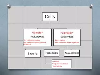
The Cell Theory • 1. All living things are composed of cells. - can be a single cell or many cells • 2. The cell is the smallest living thing that shows all of the characteristics of life. • homeostasis, metabolism, responsiveness, reproduction, evolution • 3. All cells come from preexisting living cells Presentation By: Mahendra Kandel.
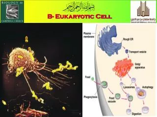
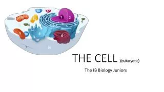
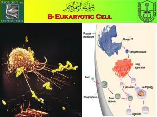
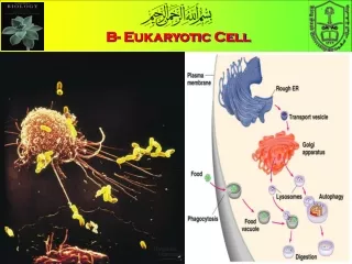
Eukaryotic Animal Cell • Eukaryotic means “true nucleus” • Eukaryotic cells have a nucleus which contains the DNA. • Eukaryotic animal cells are surrounded by a cell membrane. • Inside is the jelly like substance called cytoplasm. • Contained in the cytoplasm is the nucleus and other organellesThe other cell organelles include the endoplasmic reticulum,(rough & smooth) mitochondria, Golgi apparatus, ribosomes, lysosomes, centrioles, cilia, nucleolus. Presentation By: Mahendra Kandel.
The Human Cell • The human cell is bounded by a plasma membrane that encloses a central nucleus surrounded by cytoplasm. • Cytoplasm contains organelles, membranous structures, and a cytoskeleton • Human cells differ in size, shape and function • Human body has at least 85 different cell types Presentation By: Mahendra Kandel.
Anatomy of a typical cell Cell membrane Cytoplasm= cytosol + organelles Organelles • Smallest: • Granule cell in cerebellum: 4 μ • RBC: 5-7 μ = 0.005-0.007 mm • Largest: • Anterior horn cell in spinal cord: 135 μ • Ovum: 120 μ = 0.12 mm • Longest: • Pseudounipolar cell (toe to brainstem) Presentation By: Mahendra Kandel.
Cytosol Cytoplasm refers to the jelly-like material with organelles in it.If the organelles were removed, the soluble part that would be left is called thecytosol. It consists mainly of water with dissolved substances such as amino acids in it.
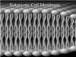
STRUCTURE This is the boundary between the cell cytoplasm & the environment Is partially permeable Made up 45% protein & 45% phospholipids with the remaining 10% cholesterol, glycoprotein & glyolipids FUNCTION Controls movement of substances in & out of the cell Forms a recognition site so that the body’s immune system can recognize its own cells Acts as a receptor site for the attachment of specific hormones and neurotransmitters. Plasma (cell) membrane Presentation By: Mahendra Kandel.
The fluid mosaic model describes the structure of the plasma membrane.Different kinds of cell membrane models have been proposed, and one of the most useful is the Fluid-mosaic model. In this model the membrane is seen as a bilayer of phospholipids in which protein molecules are embedded. An illustration of the Fluid mosaic model Presentation By: Mahendra Kandel.
Channels/pores- A channel in the cell's plasma membrane. This channel is made up of certain proteins whose function is to control the movement of food and water into the cell. These channels are made up of certain proteins. Presentation By: Mahendra Kandel.
Exocytosis Fig 2.17 Presentation By: Mahendra Kandel.
Vesicles- This term literally means "small vessel". This organelle helps store and transport products produced by the cell. The vesicles are the transport and delivery vehicles like our mail and Federal Express trucks. Some vesicles deliver materials to parts of the cell and others transport materials outside the cell in a process called exocytosis Presentation By: Mahendra Kandel.
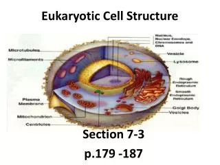
Figure 7-5 Plant and Animal Cells Animal Cell Section 7-2 Cytoplasm Nucleolus Nucleus Cell Membrane Presentation By: Mahendra Kandel. Go to Section:
Eukaryotic Cell Organelles and Function • Nucleus • Nickname: “The Control Center” • Function: holds the DNA • Parts: • Nucleolus: dark spot in the middle of the nucleus that helps make ribosomes Presentation By: Mahendra Kandel.
Nucleus- The nucleus is the control center of the cell. It is the largest organelle in the cell and it contains the DNA of the cell. The DNA of all cells is made up of chromosomes. DNA (Deoxyribonucleic Acid) contains all the information for cells to live, perform their functions and reproduce. Inside the nucleus is another organelle called the nucleolus. The nucleolus is responsible for making ribosomes. The circles on the surface of the nucleus are the nuclear pores. These are where ribosomes, and other materials move in and out of the cell. Presentation By: Mahendra Kandel.
STRUCTURE Largest organelle in the cell (10um diameter) Surrounded by a nuclear membrane / envelope Double membrane – outer is continuous with the ER Nuclear pores in the membrane allow the passage of large molecules in & out (eg messengerRNA) Material inside the nucleus is called nucleoplasm – this contains chromatin which makes up the DNA of the cell – in non-dividing cells it is spread out and during cell division it condenses to form the chromosomes A spherical structure called the nucleolus is found in the nucleus – this makes ribosomal RNA and assembles the ribosomes. FUNCTION Acts as the control centre of the cell through the production of mRNA and protein synthesis Retains the genetic material in the cell in the form of DNA / chromosomes Manufactures ribosomal RNA (rRNA) & ribosomes Starts the process of cell division Nucleus, Nucleolus & Nuclear envelope Presentation By: Mahendra Kandel.
Figure 7-5 Plant and Animal Cells Animal Cell Section 7-2 Cytoplasm Nucleolus Ribosomes Nucleus Cell Membrane Presentation By: Mahendra Kandel. Go to Section:
Eukaryotic Cell Organelles and Function • Ribosomes • Function: makes proteins • Found in all cells, prokaryotic and eukaryotic Presentation By: Mahendra Kandel.
Ribosomes- Organelles that help in the synthesis of proteins. Ribosomes are made up of two parts, called subunits. They get their names from their size. One unit is larger than than the other so they are called large and small subunits. Both these subunits are necessary for protein synthesis in the cell. When the two units are docked together with a special information unit called messenger RNA, they make proteins. Some ribosomes are found in the cytoplasm, but most are attached to the endoplasmic reticulum. While attached to the ER, ribosomes make proteins that the cell needs and also ones to be exported from the cell for work elsewhere in the body. Presentation By: Mahendra Kandel.
STRUCTURE Small dense structures found in huge numbers. Can be attached to the rough ER of floating in the cytoplasm. Are about 20 – 25 nm in diameter in eukaryotic cells and slightly smaller in prokaryotic cells Made up from two sub units FUNCTION Synthesize proteins Synthesize enzymes Ribosomes Presentation By: Mahendra Kandel.
Eukaryotic Cell Organelles and Function • Endoplasmic Reticulum (ER) • Nickname: “Roads” • Function: The internal delivery system of the cell Presentation By: Mahendra Kandel.
Figure 7-5 Plant and Animal Cells Animal Cell Section 7-2 Cytoplasm Nucleolus Ribosomes Nucleus Cell Membrane Smooth Endoplasmic Reticulum Rough Endoplasmic Reticulum Presentation By: Mahendra Kandel. Go to Section:
Endoplasmic reticulum (ER)- It is a network of membranes throughout the cytoplasm of the cell. There are two types of ER. When ribosomes are attached it is called rough ER and smooth ER when there are no ribosomes attached. The rough endoplasmic reticulum is where most protein synthesis occurs in the cell. The function of the smooth endoplasmic reticulum is to synthesize lipids in the cell. The smooth ER is also helps in the detoxification of harmful substances in the cell. Presentation By: Mahendra Kandel.
STRUCTURE Complex system of sheet like double membranes continuous with the nuclear membrane Fluid filled spaces/sacs between the membranes called CISTERNAE which allow materials to be transported through cell Two types of ER – smooth – has no ribosomes attached(SER) rough – has ribosomes attached (RER) FUNCTION Forms an extensive transport system Site of protein synthesis (Rough ER) Site of lipid, steroid and carbohydrate synthesis (smooth ER) Stores and transports these materials Endoplasmic Reticulum Presentation By: Mahendra Kandel.
Endoplasmic Reticulum • 2 Types: • Rough ER: • Rough appearance because it has ribosomes • Function: helps make proteins, that’s why it has ribosomes • Smooth ER: • NO ribosomes • Function: makes fats or lipids Presentation By: Mahendra Kandel.
Figure 7-5 Plant and Animal Cells Animal Cell Section 7-2 Cytoplasm Ribosomes Nucleolus Nucleus Cell Membrane Smooth Endoplasmic Reticulum Rough Endoplasmic Reticulum Golgi Complex Presentation By: Mahendra Kandel. Go to Section:
Eukaryotic Cell Organelles and Function • Golgi Complex • Nickname: The shippers • Function: packages, modifies, and transports materials to different location inside/outside of the cell • Appearance: stack of pancakes Presentation By: Mahendra Kandel.
Golgi complex- It is organelle in the cell that is responsible for sorting and correctly shipping the proteins produced in the ER. Just like our postal packages which should have a correct shipping address, the proteins produced in the ER, should be correctly sent to their respective address. In the cell, shipping and sorting done by the Golgi complex. It is a very important step in protein synthesis. If the Golgi complex makes a mistake in shipping the proteins to the right address, certain functions in the cell may stop. Presentation By: Mahendra Kandel.
STRUCTURE Formed from small pieces of rough ER which form small vesicles which join to make a Golgi body Chemicals made in the ER collect in the Golgi body where they are modified Small vesicles can then be ‘pinched’ off the Golgi body carrying new chemicals away which are secreted when the vesicle reaches the cell membrane Some of the vesicles become lysosomes FUNCTION Assembling glycoproteins (such as mucin) by combining carbohydrate and protein Transporting and storing lipids Formation of lysosomes Producing digestive enzymes Golgi apparatus Presentation By: Mahendra Kandel.
Figure 7-5 Plant and Animal Cells Animal Cell Section 7-2 Cytoplasm Nucleolus Ribosomes Nucleus Cell Membrane Smooth Endoplasmic Reticulum Rough Endoplasmic Reticulum Golgi Bodies Presentation By: Mahendra Kandel. Go to Section:
Eukaryotic Cell Organelles and Function • Lysosomes: circular, but bigger than ribosomes) • Nickname: “Clean-up Crews” • Function: to break down food into particles the rest of the cell can use and to destroy old cells Presentation By: Mahendra Kandel.
STRUCTURE Small vacuoles formed when small pieces of Golgi body are pinched off Contain hydrolytic enzymes which digest materials in the cell FUNCTION Release enzymes which destroy worn out organelles Digest material taken into the cell (eg white blood cells which have engulfed a bacterium) – phagocytosis Release enzymes to the outside of the cell which digest material around the cell – exocytosis Completely break down cells after they have died – autolysis Lysosomes Presentation By: Mahendra Kandel.
Figure 7-5 Plant and Animal Cells Animal Cell Section 7-2 Cytoplasm Nucleolus Ribosomes Nucleus Cell Membrane Mitochondria Smooth Endoplasmic Reticulum Rough Endoplasmic Reticulum Golgi Bodies Presentation By: Mahendra Kandel.
Eukaryotic Cell Organelles and Function • Mitochondria • Nickname: “The Powerhouse” • Function: Energy formation • Breaks down food to make ATP • ATP: is the major fuel for all cell activities that require energy Presentation By: Mahendra Kandel.
Mitochondria Mitochondria are membrane-enclosed organelles distributed through the cytosol of most eukaryotic cells. Their main function is the conversion of the potential energy of food molecules into ATP. Every type of cell has a different amount of mitochondria.. There are more mitochondria in cells that have to perform lots of work, for example- your leg muscle cells, heart muscle cells etc. Other cells need less energy to do their work and have less mitochondria. Presentation By: Mahendra Kandel.
STRUCTURE Relatively large organelle Rod/sausage shaped – 1um – 5um Have a double membrane The outer controls the entry & exit of materials Inner has many folds called cristae Surface of each crista is covered with stalked particles where ATP is made Mitochondria are filled with a jelly like matrix The matrix contains proteins, lipids, ribosomes and loops of DNA Mitochondria can replicate themselves when the cell divides FUNCTION Site of aerobic respiration (Krebs cycle & oxidative phosphorylation) Responsible for the production of energy rich ATP molecules The numbers of mitochondria reflect the metabolic activity of the cell – so large numbers are found in muscle and liver cells Mitochondria Presentation By: Mahendra Kandel.
Animal Cell Cytoplasm Nucleolus Ribosomes Nucleus Cell Membrane Mitochondria Smooth Endoplasmic Reticulum Rough Endoplasmic Reticulum Golgi Bodies Presentation By: Mahendra Kandel.
Centrosome The centrosome, also called the "microtubule organizing center", is an area in the cell where microtubles are produced. Within an animal cell centrosome there is a pair of small organelles, the centrioles, each made up of a ring of nine groups of microtubules. There are three fused microtubules in each group. The two centrioles are arranged such that one is perpendicular to the other. During animal cell division, the centrosome divides and the centrioles replicate (make new copies). The result is two centrosomes, each with its own pair of centrioles. The two centrosomes move to opposite ends of the nucleus, and from each centrosome, microtubules grow into a "spindle" which is responsible for separating replicated chromosomes into the two daughter cells. Presentation By: Mahendra Kandel.
cilia are thread-like projections of certain cells that beat in a regular fashion to create currents that sweep materials along; Presentation By: Mahendra Kandel.
Flagella may extend to the rear of a cell and push it forward by snakelike wriggling, or stick out in front and draw it along. We humans possess both flagella and cilia. Each sperm cell is propelled by a trailing flagellum that accelerates the little torpedo forward in its quest to fertilize an egg. Presentation By: Mahendra Kandel.
Some abbreviations • nm: nano metre ( 10 to the power -9 metre ) • um: micro metre ( 10 to the power -6 metre) • mm: milli metre (10 to the power -3 metre) • 1 angstrom =10 to the power -7 mm. • 1 micron (u)= 10 to the power -3 mm • 1 millimicron (mu)= 10 to the power -6mm Presentation By: Mahendra Kandel.