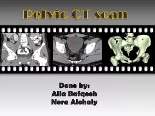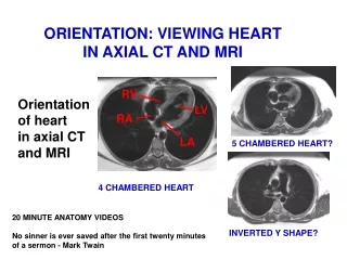
Axial Pelvic CT
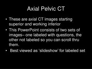
Axial Pelvic CT. These are axial CT images starting superior and working inferior This PowerPoint consists of two sets of images– one labeled with questions, the other not labeled so you can scroll thru them. Best viewed as ‘slideshow’ for labeled set. This first image is at the level

Axial Pelvic CT
E N D
Presentation Transcript
Axial Pelvic CT • These are axial CT images starting superior and working inferior • This PowerPoint consists of two sets of images– one labeled with questions, the other not labeled so you can scroll thru them. • Best viewed as ‘slideshow’ for labeled set
This first image is at the level of the aortic bifurcation and appendix (click to highlight)
Inferior Inferior vena cava Can you find the inferior vena cava? (click for answer)
What are the structures in green and blue? (please click) These are the cecum and terminal ileum
This is oral contrast (white) in loops of small bowel.
Can you find the iliac crests bilaterally? (click for answer)
Can you find the psoas muscles bilaterally? (click for answer)
Can you find the right and left Common iliac veins? (click for answer)
What are the two structures in red? (click to highlight) These are the right external andinternal Iliac arteries
Can you find the right and left iliacus muscles? (click for answer)
What is being illustrated by the dotted lines? (click to highlight) These are the Sacroiliac joints
What structures are in red bilaterally? (click to highlight) These are the Iliopsoas muscles
Can you find the left external iliac artery and vein? (click for answer)
What is the structure in yellow? (click to highlight) This is the Urinary bladder
This is the rectum What is the air filled structure (black) that is posterior to the bladder?
Can you find the piriformis muscles bilaterally? (click for answer)
What are these structures in brown? (Hint: this is a male) (click to highlight) These are the seminal vesicles
Obdurator internus muscles Can you find the obturator internus muscles bilaterally? (click for answer)
Can you find the rectus abdominus muscles? (click for answer)
What is the structure in brown? (Hint: this is a male) (click to highlight) This is the prostate
i Pubic symphisis
Inferior Inferior vena cava