Download
1 / 126
1.26k likes | 1.59k Vues
Surgical complications of cirrhosis and portal hypertension. Adam Durczyński. Normal Liver. Autopsy. 1.5 kg, wedge shape 4 lobes, Right, left, Caudate, Quadrate. Double blood supply Hepatic arteries Portal – Venous blood Acini / Portal triad. Normal Liver - Microscopy.

E N D
Surgical complications of cirrhosis and portal hypertension Adam Durczyński
Autopsy • 1.5 kg, wedge shape • 4 lobes, Right, left, Caudate, Quadrate. • Double blood supply • Hepatic arteries • Portal – Venous blood • Acini / Portal triad.
Liver Functions: • Metabolism – Carbohydrate, Fat & Protein • Secretory – bile, Bile acids, salts & pigments • Excretory – Bilirubin, drugs, toxins • Synthesis – Albumin, coagulation factors • Storage – Vitamins, carbohydrates etc. • Detoxification – toxins, ammonia, etc.
Cirrhosis was first described in a 4th century B.C. hippocratic aphorism: In cases of jaundice it is a bad sign when the liver becomes hard.
Deleteriouseffect of alcohol on the liver was appreciated by Galen and his contemporaries in the 2nd century
Alcoholic liver disease as an entity was first recognized by Baillie and other English writers after the “gin plague” in the 18th century.
Laënnec introduced the term cirrhosis, which was derived from the Greek wordkirrhos, meaning “orange-yellow.”
Etiology of Cirrhosis • Alcoholic liver disease60-70% • Viral hepatitis10% • Biliary disease (prolongedcholestasis) 5-10% • Primary hemochromatosis5% • Cryptogenic cirrhosis10-15% • Wilson’s disease • a1 –antitrypsindeficiency • Autoimmunity(lupoid hepatitis) • Cardiac cirrhosis. Due to chronic right sided heart failure which leads to liver congestion. • Galactosemia • Glycogen storage disease typu IV • Cystic fibrosis • Hepatotoxic drugs or toxins • Certain parasitic infections (such as schistosomiasis)
Introduction • Cirrhosis is common end result of many chronic liver disorders. • Diffuse scarring of liver – follows hepatocellular necrosis of hepatitis. • Inflammtion – healing with fibrosis - Regeneration of remaining hepatocytes form regenerating nodules. • Loss of normal architecture & function.
Pathogenesis: • Hepatocyte injury leading to necrosis. • Alcohol, virus, drugs, toxins, genetic etc.. • Chronic inflammation - (hepatitis). • Bridging fibrosis. • Regeneration of remaining hepatocytes proliferate as round nodules. • Loss of vascular arrangement results in regenerating hepatocytes ineffective.
Although the mechanisms are diverse, the pathologic response is uniform: hepatocellular necrosis followed by fibrosis and nodular regeneration. Each of these elements may exist alone (necrosis, uncomplicated hepatitis; fibrosis, congenital hepatic fibrosis; nodular regeneration, partial nodular transformation), but all three arerequired for the development of cirrhosis.
Normal Liver Histology CV PT
Cirrhosis Fibrosis Regenerating Nodule
Signs and symptoms • Spider angiomata or spider nevi. Vascular lesions consisting of a central arteriole surrounded by many smaller vessels due to an increase in estradiol. These occur in about 1/3 of cases. • Palmar erythema. Exaggerations of normal speckled mottling of the palm, due to altered sex hormone metabolism.
Nail changes. • Muehrcke's nails - paired horizontal bands separated by normal color due to hypoalbuminemia (low production of albumin). • Terry's nails - proximal two thirds of the nail plate appears white with distal one-third red, also due to hypoalbuminemia • Clubbing - angle between the nail plate and proximal nail fold > 180 degrees
Hypertrophic osteoarthropathy. Chronic proliferative periostitis of the long bones that can cause considerable pain. • Dupuytren's contracture. Thickening and shortening of palmar fascia that leads to flexion deformities of the fingers. Thought to be due to fibroblastic proliferation and disorderly collagen deposition. It is relatively common (33% of patients). • Gynecomastia. Benign proliferation of glandular tissue of male breasts presenting with a rubbery or firm mass extending concentrically from the nipples. This is due to increased estradiol and can occur in up to 66% of patients.
Jaundice. Yellow discoloring of the skin, eye, and mucus membranes due to increased bilirubin (at least 2–3 mg/dL or 30 mmol/L). Urine may also appear dark.
Caput medusa. In portal hypertension, the umbilical vein may open. Blood from the portal venous system may be shunted through the periumbilical veins into the umbilical vein and ultimately to the abdominal wall veins, manifesting as caput medusa.
Ascites. Accumulation of fluid in the peritoneal cavity giving rise to flank dullness (needs about 1500 mL to detect flank dullness). It may be associated with hydrocele and penile flomation (swelling of the penile shaft) in men.
Hypogonadism. Manifested as impotence, infertility, loss of sexual drive, and testicular atrophy due to primary gonadal injury or suppression of hypothalamic or pituitary function. • Liver size. Can be enlarged, normal, or shrunken. • Splenomegaly (increase in size of the spleen). Due to congestion of the red pulp as a result of portal hypertension. • Cruveilhier-Baumgarten murmur. Venous hum heard in epigastric region (on examination by stethoscope) due to collateral connections between portal system and the remnant of the umbilical vein in portal hypertension. • Fetor hepaticus. Musty odor in breath due to increased dimethyl sulfide. • Asterixis. Bilateral asynchronous flapping of outstretched, dorsiflexed hands seen in patients with hepatic encephalopathy. • Other. Weakness, fatigue, anorexia, weight loss.
Complications • As the disease progresses, complications may develop. In some people, these may be the first signs of the disease. • Bruising and bleeding due to decreased production of coagulation factors. • Jaundice due to decreased processing of bilirubin. • Itching (pruritus) due to bile products deposited in the skin. • Hepatic encephalopathy - the liver does not clear ammonia and related nitrogenous substances from the blood, which are carried to the brain, affecting cerebral functioning: neglect of personal appearance, unresponsiveness, forgetfulness, trouble concentrating, or changes in sleep habits. • Sensitivity to medication due to decreased metabolism of the active compounds. • Hepatocellular carcinoma is primary liver cancer, a frequent complication of cirrhosis. It has a high mortality rate.
Portal hypertension - blood normally carried from the intestines and spleen through the hepatic portal vein flows more slowly and the pressure increases; this leads to the following complications: • Ascites - fluid leaks through the vasculature into the abdominal cavity. • Esophageal varices - collateral portal blood flow through vessels in the stomach and esophagus. These blood vessels may become enlarged and are more likely to burst. • Problems in other organs. • Cirrhosis can cause immune system dysfunction, leading to infection. Signs and symptoms of infection may be aspecific are more difficult to recognize (e.g. worsening encephalopathy but no fever). • Fluid in the abdomen (ascites) may become infected with bacteria normally present in the intestines (spontaneous bacterial peritonitis). • Hepatorenal syndrome - insufficient blood supply to the kidneys, causing acute renal failure. This complication has a very high mortality (over 50%). • Hepatopulmonary syndrome - blood bypassing the normal lung circulation (shunting), leading to cyanosis and dyspnea (shortness of breath), characteristically worse on sitting up. • Portopulmonary hypertension - increased blood pressure over the lungs as a consequence of portal hypertension.
Lab findings The following findings are typical in cirrhosis: • Aminotransferases - AST and ALT are moderately elevated, with AST > ALT. However, normal aminotransferases do not preclude cirrhosis. • Alkaline phosphatase - usually slightly elevated. • GGT – correlates with AP levels. Typically much higher in chronic liver disease from alcohol. • Bilirubin - may elevate as cirrhosis progresses. • Albumin - levels fall as the synthetic function of the liver declines with worsening cirrhosis since albumin is exclusively synthesized in the liver • Prothrombin time - increases since the liver synthesizes clotting factors. • Globulins - increased due to shunting of bacterial antigens away from the liver to lymphoid tissue. • Serum sodium - hyponatremia due to inability to excrete free water resulting from high levels of ADH and aldosterone. • Thrombocytopenia - due to both congestive splenomegaly as well as decreased thrombopoietin from the liver. However, this rarely results in platelet count < 50,000/mL.
Leukopenia and neutropenia - due to splenomegaly with splenic margination. • Coagulation defects - the liver produces most of the coagulation factors and thus coagulopathy correlates with worsening liver disease. • Other laboratory studies performed in newly diagnosed cirrhosis may include: • Serology for hepatitis viruses, autoantibodies (ANA, anti-smooth muscle, anti-mitochondria, anti-LKM) • Ferritin and transferrin saturation (markers of iron overload), copper and ceruloplasmin (markers of copper overload) • Immunoglobulin levels (IgG, IgM, IgA) - these are non-specific but may assist in distinguishing various causes • Cholesterol and glucose • Alpha 1-antitrypsin
Measurement of Hepatic Functional ReserveChild-Pugh Criteria for Hepatic Functional Reserve
The portal vein is formed from the confluence of the superior mesenteric and splenic veins behind the neck of the pancreas and is 6 to 8 cm in length. The left gastric or coronary vein drains the distal esophagus and lesser curvature of the stomach, generally entering the portal vein near its origin. The splenic vein lies beneath the pancreas and is usually joined by the inferior mesenteric vein just before its confluence with the superior mesenteric vein.
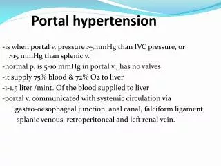
Portal hypertension • Portal pressure gradient 12 mmHg or more • Often associated with varices and ascites. • Many conditions are associated with it, the most common being cirrhosis of the liver.
Pathophysiology • P=FR, where P is pressure gradient thru the portal system, F is the volumeof blood flowing thru the system, R is the resistance to flow. • Changes in either F or R affect the pressure. • In most types of portal hypertension, both flow and resistance are altered.
Increase in resistance • Liver disease is responsible for a decrease in portal vascular radius, producing an increase in portal vascular resistance. • In cirrhosis, the increase occurs at the microcirculation (sinusoidal). • The resistance is also due to active myofibroblasts, vascular smooth muscle cells in the intrahepatic veins.
Increase in Flow • The increase in blood flow is caused by splanchnic arteriolar vasodilitation caused by release of endogenous vasodilators. • The increased flow aggravates the increase in portal pressure and contributes to why PT exists despite the formation of portosystemic collaterals that divert as much as 80% of portal flow.
Imaging Studies • Duplex is safe, noninvasive. Demonstrates portal flow, portal vein thrombosis, splenic vein thrombosis • Nodular liver surface, splenomegaly, presence of collateral circulation. • Limitations include meals, meds, sympathetic nervous system affect flow.
Imaging Studies • CT scan when US inconclusive • Look for collaterals from portal system • Dilatation of the vena cava suggests portal hypertension. • Limitations include not being able to use IV contrast in allergic patients or with renal failure.
Procedures • Hemodynamic measurement of pressure, usually not performed due to invasive nature. Measures hepatic venous pressure gradient (HVPG). Similar to Swan Ganz, where balloon is inflated measuring wedged hepatic venous pressure, minus the unoccluded pressure is the HVPG.
Procedures • Endoscopy is performed to screen for varices. • Gastroesophageal varices confirms diagnosis of portal hypertension, absence does not rule it out. • Many times an incidental finding when scoped for something else.
Pathogenesis Varices in the distal esophagus and proximal stomach are a component of the collateral network that diverts high-pressure portal venous flow through the left and rightgastric veins and the short gastric veins to the azygous system. Esophagogastric varices do not bleed until portal pressure exceeds 12 mm Hg, and then they bleed in only one third to one half of patients. The three key variables that are predictive of variceal bleeding are Child-Pugh class, variceal size, and the presence and severity of red wale markings (indicative of epithelial thickness).