
Understanding Epithelial Tissue: Functions, Types & Characteristics
E N D
Presentation Transcript
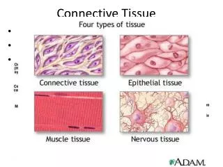
Tissue • Tissue - A group of closely associated cells that are similar in structure and perform related functions • Four primary types • Epithelium • Connective tissue • Nervous tissue • Muscle
Tissue • Epithelium: epithe = laid on, covering
Epithelial Tissue • Boundary: covers body surface or lines a body cavity (i.e.: digestive and respiratory) • Forms parts of most glands *Nearly all substances given or received by the body must pass through epithelium
Epithelial Tissue • Functions of epithelium • Protection: body’s first line of defense • Absorption – gas exchange • Ion transport • Filtration • Secretion – sweat, digestive juices, mucous, lubricant • Sensory input: skin, nose, eyes, ears
Special Characteristics of Epithelia CELLS, CELLS, CELLS • Cellularity –close/tight fit cells, forming sheets, bound at many points & separated by minimal extra cellular material • Specialized contacts - esp tight j’ns & desmosomes • Polarity - cell regions of the apical surface differ from the basal surface
Special Characteristics of Epithelia • Polarity • Apical surface - unattached, exposed to body’s exterior or organ cavity – often slick or covered w/cilia or villi • Basal surface – anchors to basement membrane
Special Characteristics of Epithelia 3 layers of basement membrane • Secreted by both epithelium & connective tissue • Functions • Filters molecules from capillaries entering epithelium • Anchors epithelium to underlying connective tissue • Non-cellular • Lamina lucida + Lamina densa = Basal lamina • Extracellular matrix composition varies: clear, delicate, structureless sheet, includes proteins • Lamina reticularis • Collagenous/protein fibers
Special Characteristics of Epithelia Basal surface/Basement membrane epithelia Lamina lumina Basal lamina Lamina densa Reticular fibers Lamina reticularis Anchoring fibers Anchoring plaque
Apical surface features • Microvilli – fingerlike extensions of plasma membrane • Abundant in small intestine and kidney • Maximize surface area across which small molecules enter or leave • Act as stiff knobs that resist abrasion
Cilia • Whiplike, highly motile extensions of apical surface membranes • Microtubules in cilia – arranged similarly to cytoplasmic organelles called centrioles • Movement of cilia – in coordinated waves Figure 4.8
Special Characteristics of Epithelia • Support by connective tissue • Avascularnutrients diffuse from underlying connective tissue • Innervated infiltrated with nerves • Regeneration - lost cells are quickly replaced by cell division if well nourished
Classifications of Epithelia • Classification includes 2 names (except transitional) • First name of tissue indicates number of cell layers • Simple – one layer of cells • Stratified – more than one layer of cells (named for cells at free/apical surface)
Classifications of Epithelia • Last name describes cell shape • Squamous (“scale-” or plate-like) • Hexagonal cells are wider than tall • Nuclei: central & flat (bumps in cross section) • Very little cytoplasm
Classifications of Epithelia • Last name of tissue describes shape of cells • Cuboidal • Cells are as wide as tall • Nuclei: large, central, round • May have microvilli, cilia
Classifications of Epithelia • Last name of tissue describes shape of cells • Columnar • Cells are taller than they are wide • Round to oval nuclei, near base • May have microvilli, cilia
Unicellular Exocrine Glands: Goblet Cells • Goblet cells - unicellular exocrine glands that produce mucin • Mucin + water mucus • Protects and lubricates many internal body surfaces • May be embedded in epithelial tissue
Simple Squamous Epithelium • Description – single layer, flat cells • Function • Diffusion • Filtration • Friction reduction: secretes lubricating substances in serosae • Location • Endothelium: Lines heart, blood & lymph vessels • Mesothelium: Lines serous membranes - peritoneal, pleural, pericardial cavities • Covers visceral organs (3 major body cavities) • Small ducts, alveoli, loops of Henle (kidney) • Inner surface of eardrum
Simple Cuboidal Epithelium • Description - single layer of cube-like cells • Function - secretion and absorption • Location – kidney tubules, secretory portions of small glands & ducts (thyroid, salivary, pancreas), ovary surface
Simple Columnar Epithelium • Description – single layer of column-shaped, some w/microvilli, goblet cells • Function • Absorption • Secretion of mucus, enzymes, and other substances • Ciliated type propels mucus or reproductive cells by ciliary action
Simple Columnar Epithelium • Nonciliated - digestive tract [stomach (protects stomach from acid) to anus], gallbladder, ducts of some glands • Microvilli (ABSORB!) & goblet cells (secrete mucous)
Simple Columnar Epithelium • Ciliated form - • In respiratory tract, goblet cells are interspersed among ciliated columnar epithelia of bronchi • Secreted mucus on the surface traps inhaled foreign particles. Beating cilia moves particles to the throat for removal by coughing, swallowing, or sneezing • Cilia also moves oocytes to the uterine tubes
Simple Columnar Epithelium • Ciliated form - small bronchi, fallopian tubes
Pseudostratified Columnar Epithelium • Description • All cells originate at basement membrane, but only tall cells reach the apical surface • May contain goblet cells and bear cilia • Nuclei lie at varying heights within cells, giving false impression of stratification
Pseudostratified nonciliated columnar epithelium • Nonciliated – no cilia, no goblet cells • Location: ducts of epididymis, vas deferens, urethra, ducts of large glands
Pseudostratified Ciliated Columnar Epithelium • Ciliated (MOST) – cilia & goblet cells • Location: lines trachea and most of upper respiratory tract
Stratified Epithelia • Properties • Regenerate from basal layer • Major role is protection – more layers
Stratified Squamous Epithelium • Description • Most common stratified epithelial tissue • Thickest epithelial tissue, adapted for protection from abrasion • Base layers are metabolically active • Deeper layers of cells appear cuboidal or columnar • Cells dehydrate (become squamous), harden, die as pushed toward apical surface
Stratified Squamous Epithelium • Two types • Keratinized – forms epidermis, surface cells are dead and full of keratin, a protective protein, waterproof • Nonkeratinized - forms moist lining of body openings of mouth, esophagus, rectum, vagina,
Stratified Squamous Epithelium Figure 4.3e
Stratified Squamous Epithelium Figure 4.3e
Stratified Squamous Epithelium Figure 4.3e
Stratified Cuboidal Epithelium • Description – rare in humans; generally two layers of cube-shaped cells • Function – protection, secretion • Location • Egg-producing vesicles (ovaries) • Sperm -producing ducts (seminiferous tubules) • Forms ducts of • Mammary glands • Salivary glands • Largest sweat glands
Stratified Cuboidal Epithelium Figure 4.3f
Stratified Cuboidal Epithelium Figure 4.3f
Stratified Cuboidal Epithelium Figure 4.3f
Stratified Cuboidal Epithelium Figure 4.3f