Download
1 / 47
650 likes | 1.56k Vues
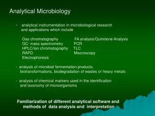
Analytical Microbiology. analytical instrumentation in microbiological research and applications which include Gas chromatography FA analysis/Quinolone Analysis GC- mass spectrometry PCR HPLC/Ion chromatography TLC

E N D
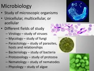
Analytical Microbiology • analytical instrumentation in microbiological research • and applications which include • Gas chromatography FA analysis/Quinolone Analysis • GC- mass spectrometry PCR • HPLC/Ion chromatography TLC • RAPD Miscroscopy • Electrophoresis • analysis of microbial fermentation products, • biotransformations, biodegradation of wastes or heavy metals • - analysis of chemical markers used in the identification • and taxonomy of microorganisms Familiarization of different analytical software and methods of data analysis and interpretation
Anton van Leeuwenhoek (1673) - using a simple microscope, he was the first to observe microorganisms. Animalcule
Leeuwenhoek’s drawings of bacteria A, C, F and G are rod –shaped E – spherical or coccus-shaped, H cocci packets
Hooke’s observation laid the groundwork for development of the cell theory, the concept that all living things are composed of cells. Cell walls in cork tissue Born on July 18, 1635 Robert Hooke (1665) - observed that plant material was composed of little boxes, he introduced the term “ cell.”
Drawing of Robert Hooke, which represents one of the first microscopic descriptions of microorganisms A blue mold growing on the surface of leather, the round structures contain spores of the mold
Microscope = 2 Greek words mikros = small skopein = to look through Three lenses/light 1. Ocular/eyepiece 2. Objectives 3. Condenser LPO = 10x (100) HPO = 40/45x (450) OIO = 100x (1000) Scanner = 4x (optional)
Microscope must accomplish 3 tasks: • produce a magnified image of the specimen • separate the details in the image • and render the details visible to the human eye or camera Together, the optical and mechanical components of the microscope, including the mounted specimen on a glass micro slide and coverslip, form an optical train with a central axis that traverses the microscope base and stand
Dissecting microscope Compound microscope Coxial binocular microscope Inverted microscope Microscope with cleaning kit Inclined microscope Research projection microscope Zoom tinocular microscope Projection microscope
Resolving Power - measures the ability to distinguish small objects close together 0.61 (lambda) r.p. = ______________ (N sinØ) where lambda = wavelength of illuminating light • for light scope, can improve R.P. by making lambda smaller or sinØ larger
R.P. is smallest for violet light, human eye is more sensitive to blue, optimal R.P. is achieved with blue light (450 nm). n sinΦ is called numerical aperture (it measures how much light cone spreads out between condenser and specimen). more spread = better resolution Φ = angle of light cone maximum value is 1.0 n = refractive index n = 1.0 in air can increase with certain oils (up to 1.4), called immersion oil N.A. is property of lens Theoretical limit of R.P. for light scope is 0.2 micrometers
Advantages: convenient, relatively inexpensive, widely available • Disadvantages: resolving power 0.2 micrometers at best can recognize cells but not fine details needs contrast; cells are mainly water and don't contrast with their medium Easiest way to view cells is to fix and stain Fixation preserves cells; disrupts proteins, prevents decay/ degradation • typical treatments: heat, formalin, glutaraldehyde Staining Simple Stains adds colored compounds -----contrast Bright-field microscope basic dyes: e.g. methylene blue, crystal violet. Cations ( + charges) bind to - charge groups on proteins, nucleic acids acidic dyes: e.g. eosin, acid fuchsin. Anions ( - charges); bind to + charges on proteins, phospholipids • Differential Stains allow differentiation between different organisms • Examples: Gram stain Spore stain
Cells are mostly water, very little contrast from surrounding medium, so not very visible in light Phase scope converts slight differences in refractive index and cell density into variations Scope uses annular stop below condenser: thin transparent ring in opaque disk ----- hollow light cone Phase contrast microscope As light passes through specimen, some rays are deviated and retarded by ¼ wavelength Have phase plate in objective lens: transparent optical disk with phase ring Undeviated light passes through ring, is advanced by ¼ wavelength bright background Deviated light doesn’t pass through phase ring, is not advanced. When light gets focused, deviated rays cancel out with undeviated rays, producing dark image where objects were Advantage: can see live material without staining
Fluors are chemicals that adsorb light to produce excited electrons, later reradiate light = fluorescence Fluorescence microscopy • - need filters to remove this light from light traveling to ocular lens • only fluoresced light emitted from object will then appear to eye • need dark field condenser to create dark background • can couple flour to specific probe molecules (usually antibodies) bind to • preparation • if sample is illuminated with wavelength of exciting light, then filter out that • wavelength to prevent reaching the sample and nothing is seen • but if fluorescence occurs, different wavelength of light is produced, object is • seen • good technique to detect specific microbe in complex sample. (e.g. detect • gonococcus in vaginal smear) • - requires correct microscope, fluors, technical skill
Differential Interference Contrast (DIC) Microscopy Uses a polarizer to create two distinct beams of polarized light Gives structures such as endospores, vacuoles and granules a three- dimensional appearance Structures not visible using bright- field microscopy are sometimes visible using DIC
The atomic force microscope (AFM) or scanning force microscope (SFM) is a very high-resolution type of scanning probe microscopy, with demonstrated resolution of fractions of a nanometer, more than 1000 times better than the optical diffraction limit. developed by Gerd Binnig and Heinrich Rohrer in the early 1980s, a development that earned them the Nobel Prize for Physics in 1986 is one of the foremost tools for imaging, measuring and manipulating matter at the nanoscale
Confocal Scanning Laser Microscopy Uses a computerized microscope coupled with a laser source to generate a three-dimensional image Computer can focus the laser on single layers of the specimen Different layers can then be compiled for a three-dimensional image Resolution is 0.1 um for CSLM
RELATIONSHIP OF STRUCTURE TO FUNCTION SIZE the difference between an average bacterium (measured in mm) and an elephant (measured in meters) V = 4/3 r3 Volume of sphere/ coccus V = r2 h Volume of cylinder/ bacillus smallest bacteria (e.g., mycoplasmas) 0.2 mm in diameter--- V = 4.18 x 10⁻¹⁵ cm3 largest bacterium known (60 mm x 600 mm--- V = 1.74 x 10⁻⁶ cm3 The ratio of these two numbers is 4.2 x 10⁸ !!! Eucaryotes span an even larger range of about 1018!!
Surface of cylinder S = 2 r h, hence S/ V = 2/r Surface of sphere S = 4 r h, hence S/ V = 3/r As r gets smaller and smaller, S/ V gets larger and larger -- difference between sphere and cylinder becomes insignificant If elephant is approximated by a sphere of 3 m, then 3/3 = 1.0 m-1 Correspondingly, -- a small mycoplasm would have S/ V = 3/1 x 10-7 = 3 x 10⁷ m-1
Methods for Measurement of Cell Mass • Direct physical measurement of dry weight, wet weight, or • volume of cells after centrifugation. 2. Direct chemical measurement of some chemical component of the cells such as total N, total protein, or total DNA content. 3. Indirect measurement of chemical activity such as rate of O2 production or consumption, CO2 production or consumption, etc. 4. Turbidity measurements employ a variety of instruments to determine the amount of light scattered by a suspension of cells. Particulate objects such as bacteria scatter light in proportion to their numbers. The turbidity or optical density of a suspension of cells is directly related to cell mass or cell number, after construction and calibration of a standard curve. The method is simple and nondestructive, but the sensitivity is limited to about 107 cells per ml for most bacteria.
Methods for Measurement of Cell Numbers 1. Direct microscopic counts are possible using special slides known as counting chambers. Dead cells cannot be distinguished from living ones. Only dense suspensions can be counted (>107 cells per ml), but samples can be concentrated by centrifugation or filtration to increase sensitivity. 2. Electronic counting chambers count numbers and measure size distribution of cells. Such electronic devices are more often used to count eukaryotic cells such as blood cells. 3. Indirect viable cell counts, also called plate counts, involve plating out (spreading) a sample of a culture on a nutrient agar surface.
Micrometry - measurement of minute objects with a micrometer.
Calibration Factor - actual distance between any two adjacent lines of the ocular micrometer by observing how many lines of the stage micrometer (Sm) are included within a given number of lines on the ocular micrometer (Om). The distance between any two adjacent lines on the stage micrometer is = 0.01 mm (10 microns) C. F. = Sm x 0.01 mm (10 microns) Om Example : 10 divisions in Om match with 6 divisions in Sm 6 x 0.01 mm = 0.006 mm 10 or 6 x 10 µm= 6µm 10
10-1 = deci 10-2 = centi 10-3 = milli 10-6 = micro 10-9 = nano 10-12 = pico 10-15 = femto 10-18 = atto • = deca • 102 = hecto • 103 = kilo • 106 = mega • 109 = giga • 1012 = tetra
The Sleeve does not move. It looks like a ruler with ten numbers. The space between each number is divided into quarters. As the Thimble rotates around this Sleeve it covers up, or reveals the numbers marked on the Sleeve.
It is easy to read a micrometer if you think of the markings on the Sleeve as dollars and quarters. What are the readings on the micrometers as shown?
As the thimble rotates, you add those pennies to the dollars and quarters
References: Brock Biology of Microorganisms Internet Sources (especially most of the diagrams)