Download
1 / 123
1.24k likes | 1.38k Vues
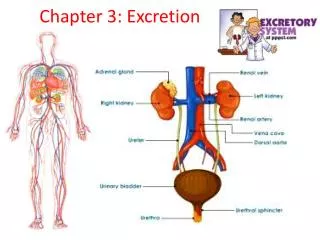
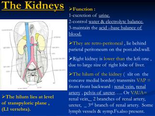
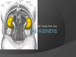
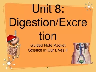
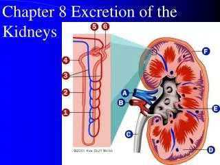
Chapter 8 Excretion of the Kidneys. Major Functions of the Kidneys 1. Regulation of: body fluid osmolarity and volume electrolyte balance acid-base balance blood pressure 2. Excretion of metabolic products foreign substances (pesticides, chemicals etc.)

E N D
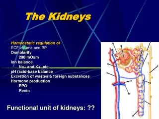
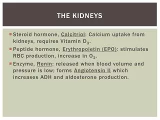
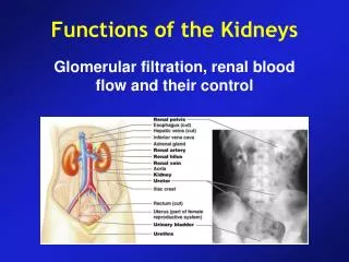
Major Functions of the Kidneys 1. Regulation of: body fluid osmolarity and volume electrolyte balance acid-base balance blood pressure 2. Excretion of metabolic products foreign substances (pesticides, chemicals etc.) excess substance (water, etc) 3. Secretion of erythropoitin 1,25-dihydroxy vitamin D3 (vitamin D activation) renin prostaglandin
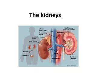
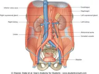
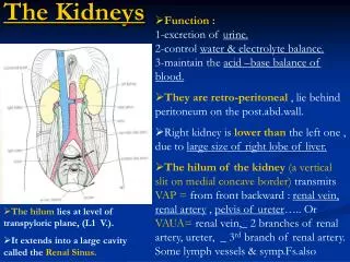
Section 1 Characteristics of Renal Structure and Function • Physiological Anatomy of the Kidney
Nephron and • Collecting Duct • Nephron: The functional unit of the kidney • Each kidney is made up of about 1 million nephrons • Each nephrons has two major components: • A glomerulus • A long tube
Cortical nephron Juxtamedullary nephron
Anatomy of Kidney • Cortical nephron • 80%-90% • glomeruli in outer cortex • short loops of Henle • extend only short distance into medulla • blood flow through cortex is rapid • cortical interstitial fluid 300 mOsmolar
Anatomy of Kidney • Juxtamedullary nephron • glomeruli in inner part of cortex • long loops of Henle • extend deeply into medulla. • blood flow through vasa recta in medulla is slow • medullary interstitial fluid is hyperosmotic • maintains osmolality, filtering blood and maintaining acid-base balance
2. The juxtaglomerular apparatus Including macula densa, extraglumerular mesangial cells, and juxtaglomerular (granular cells) cells
3. Characteristics of the renal blood flow: 1, High blood flow. 1200 ml/min, or 21 percent of the cardiac output. 94% to the cortex 2, Two capillary beds High hydrostatic pressure in glomerular capillary (about 60 mmHg) and low hydrostatic pressure in peritubular capillaries (about 13 mmHg) Vesa Recta
Functions of the Nephron Secretion Reabsorption Excretion Filtration
Filtration • First step in urine formation • Bulk transport of fluid from blood to kidney tubule • Isosmotic filtrate • Blood cells and proteins don’t filter • Result of hydraulic pressure • GFR = 180 L/day
Reabsorption • Process of returning filtered material to bloodstream • 99% of what is filtered • May involve transport proteins • Normally glucose is totally reabsorbed
Secretion • Material added to lumen of kidney from blood • Active transport (usually) of toxins and foreign substances • Saccharine (糖精) • Penicillin
Excretion: • Loss of fluid from body in form of urine • Amount = Amount + Amount -- Amount • of Solute Filtered Secreted Reabsorbed • Excreted
Glomerular filtration • blood enters glomerular capillary • filters out of renal corpuscle • large proteins and cells stay behind • everything else is filtered into nephron • glomerular filtrate • plasma like fluid
Factors that determining the glumerular filterability • Molecular weight • Charges of the molecule
Filtration Membrane • One layer of glomerular capillary cells. • Fenestration, 70 – 90 nm, permeable to protein of small molecular C: capillary F: fenestration BM: basal membrane P podocytes FS: filtration slit
Filtration Membrane • Basement membrane(lamina densa) • with the mesh of 2-8 nm diameter C: capillary F: fenestration BM: basal membrane P podocytes FS: filtration slit
Filtration Membrane • One layer of cells in Bowman’s capsule: • Podocytes have foot like projections (pedicels) with filtration slits (滤过裂隙)in between C: capillary F: fenestration BM: basal membrane P podocytes FS: filtration slit
Dextran filterability 右旋糖苷 Stanton BA & Koeppen BM:‘The Kidney’ in Physiology,Ed. Berne & Levy, Mosby, 1998 2934
Protein filtration: influence of negative charge on glomerular wall
Starling Forces Involved in Filtration: What forces favor/oppose filtration?
Glomerular filtration • Mechanism: Bulk flow • Direction of movement : From glomerular capillaries to capsule space • Driving force: Pressure gradient (net filtration pressure, NFP) • Types of pressure: • Favoring Force: Capillary Blood Pressure (BP), Opposing Force: Blood colloid osmotic pressure(COP) and Capsule Pressure (CP)
Glomerular filtration rate (GFR) • Amount of filtrate produced in the kidneys each minute. 125mL/min = 180L/day • Factors that alter filtration pressure change GFR. These include: • Increased renal blood flow -- Increased GFR • Decreased plasma protein -- Increased GFR. Causes edema. • Hemorrhage -- Decreased capillary BP -- Decreased GFR • Capsular pressure
GFR regulation : Adjusting blood flow • GFR is regulated by three mechanisms • 1. Renal Autoregulation • 2. Neural regulation • 3. Hormonal regulation • All three mechanism adjust renal blood pressure and resulting blood flow
1. Renal autoregulation ERPF: experimental renal plasma flow GFR: glomerular filtration rate
Mechanism? • Myogenic Mechanism • Tubuloglomerular feedback
1) Myogenic Mechanism of the autoregulation Blood Flow = Capillary Pressure / Flow resistance
2. Neural regulation of GFR • Sympathetic nerve fibers innervate afferent and efferent arteriole • Normally sympathetic stimulation is low but can increase during hemorrhage and exercise
3. Hormonal regulation of GFR • Angiotensin II. • a potent vasoconstrictor. • Reduces GFR • ANP (Atrial Natriuretic Peptide) • increases GFR by relaxing the afferent arteriole NO • Endothelin • Prostaglandin E2
Measuring GFR • 125ml/min, 180L/day • plasma clearance: • The amount of a kind of substance present in urine • The substance: filtered but neither reabsorbed nor secreted, • If plasma conc. is 3mg/L then • 3mg/L X 180/day = 540mg/day • (known) (unknown) (known)
Renal handling of inulin菊粉 Amount filtered = Amount excreted Pin x GFR Uin x V
Qualities of agents to measure GFR • Inulin: (Polysaccharide from Dahalia plant) • Freely filterable at glomerulus • Does not bind to plasma proteins • Biologically inert • Non-toxic, neither synthesized nor metabolized in kidney • Neither absorbed nor secreted • Does not alter renal function • Can be accurately quantified • Low concentrations are enough (10-20 mg/100 ml plasma)
Qualities of agents to measure GFR Creatinine (肌氨酸酐): End product of muscle creatine (肌氨酸) metabolism Used in clinical setting to measure GFR but less accurate than inulin method Small amount secreted from the tubule
Section 3 Reabsorption and Secretion Concept of Reabsorption and Secretion
GFR 125 ml/min (180L/day) • (about 1% is excreted)
Filtration, reabsoption, and excretion rates of substances by the kidneys Filtered Reabsorbed Excreted Reabsorbed (meq/24h) (meq/24h) (meq/24h) (%) Glucose (g/day) 180 180 0 100 Bicarbonate (meq/day) 4,320 4,318 2 > 99.9 Sodium (meq/day) 25,560 25,410 150 99.4 Chloride (meq/day) 19,440 19,260 180 99.1 Water (l/day) 169 167.5 1.5 99.1 Urea (g/day) 48 24 24 50 Creatinine (g/day) 1.8 0 1.8 0
Two pathways of the absorption: Transcellular Pathway Paracellular transport Plasma Lumen Cells
Mechanism of Transport 1, Primary Active Transport 2, Secondary Active Transport 3, Pinocytosis 4, Passive Transport
Secondary active transport Tubular lumen Interstitial Fluid Interstitial Fluid Tubular lumen Tubular Cell Tubular Cell co-transport counter-transport (symport) (antiport) out in out in Na+ Na+ glucose H+ Co-transporters will move one moiety, e.g. glucose, in the same direction as the Na+. Counter-transporters will move one moiety, e.g. H+, in the opposite direction to the Na+.
Passive Transport Diffusion