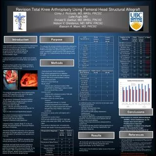
Left Atrial Volume Index During Diastasis as a Predictor of Adverse Cardiac Events
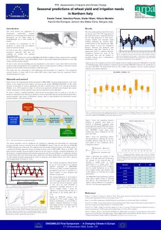
This study investigates the prognostic value of Left Atrial Volume Index (LAVI) assessed during ventricular diastasis using cardiac computed tomography (CT). By analyzing data from 101 patients with adverse events and a matched control group, we found that LAVI is significantly larger in those experiencing adverse events, highlighting its incremental predictive ability. The findings suggest that LAVI provides essential prognostic information that can enhance patient assessment during non-invasive CAD evaluations while minimizing radiation exposure, potentially impacting clinical decision-making.

Left Atrial Volume Index During Diastasis as a Predictor of Adverse Cardiac Events
E N D
Presentation Transcript
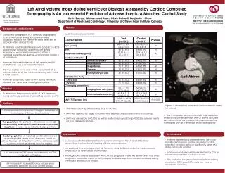
Left Atrial Volume Index during Ventricular Diastasis Assessed by Cardiac Computed Tomography is An Incremental Predictor of Adverse Events: A Matched Control Study Kevin Boczar, Mohammed Alam, Girish Dwivedi, Benjamin J Chow Department of Medicine (Cardiology), University of Ottawa Heart Institute, Canada , Results Background and Rationale • Table: Baseline characteristics • Computed tomographic (CT) coronary angiography (CTA) is increasingly being accepted as a key diagnostic modality for the non-invasive detection of coronary artery disease (CAD) • To minimize patient radiation exposure prospective-ECG gated image acquisition algorithms are being increasingly used whereby image acquisition is restricted to ventricular diastasis when cardiac motion is at a minimum. • However, this leads to the loss of left ventricular (LV) and left atrial (LA) functional information. • Previous studies have shown that assessment of LA volume index (LAVI) has incremental prognostic value in CAD patients. • However, prognostic value of LAVI during ventricular diastasis has never been investigated before. Left Atrium Objective • To determine the prognostic ability of LAVI assessed during ventricular diastasis in predicting adverse events Methods Figure : 3 dimensional volumetric method used to assess LA volume • The mean follow up duration was 20 ± 12 months. • LAVI was significantly larger in patients who experienced adverse events on follow up. • LAVI was univariable (p=0.001) as well as multivariable predictor (p=0.001) of adverse events on Cox regression analysis Patients recruited from Cardiac CT registry Database at the University of Ottawa Heart Institute • True 3 dimension reconstruction with high resolution endocardial border definition with CT allows accurate estimation of LAV not possible with other competitive techniques such as 2 dimension echocardiography. Test population: 101 patients with adverse events (all-cause mortality and troponin positive acute myocardial infarction) on follow- up constituted test population. Conclusions Discussion Control population: .A matched control list (matched according to the Morise score: the score based on clinical findings ) of 101 patients with no adverse events on follow-up was generated from the registry. • Patients experiencing adverse events (all-cause mortality and troponin positive acute myocardial infarction) on follow-up have significantly larger LAVI during ventricular diastasis . • LAVI assessed during ventricular diastasis by CT is an incremental predictor of adverse events. • This additional prognostic information from existing prospective ECG-gated CTA data sets may be provided to clinicians. • . • LA is exposed to the (diastolic) haemodynamic changes in the LV due to the close anatomical and functional coupling of these two chambers. • An enlarged LA is an independent risk factor for atrial fibrillation and other cardiovascular events such as heart failure and mortality. • Although CAD severity assessment with CTA has prognostic value, we demonstrate that other prognostic information (such as LAVI) may be available even from datasets obtained during ventricular diastasis (75% phase). LAV measured at 75% phase (mid-diastole) on CTA imaging in both groups LAV Indices (LAV indexed to body surface area) compared between test and control groups