
Understanding Hernias: Causes, Types, and Treatments
E N D
Presentation Transcript
Hernias Professor Oseremen Aisuodionoe- Shadrach
Introduction • Hernia is a protrusion of a viscus or part of a viscus through an abnormal opening in the walls of its containing cavity • One of the most common condition in surgical practice • Accounts for up to 75% of the causes of intestinal obstruction
Aetiology • All hernias occur at the sites of congenital weakness or potential weakness of the abdominal wall which are acted on by a continued or repeated increase in abdominal pressure • such sites are • where blood vessels and other structures enter or leave the abdominal or thoracic cavity • Muscle fail to overlap • No muscle, only scar tissue e.g umbilicus • Acquired e.g. post surgery
Predisposing factors • Congenital defects: A congenital peritoneal sac predispose to hernia formation in early life and can result in • Persistence of processus vaginalis- indirect inguinal hernia • Incomplete obliteration of umbilicus- umbilical hernia • Patient canal of Nück- indirect inguinal hernia formation in females
Persistent communication between abdominal and thoracic cavity – Diaphragmatic Hernia • Acquired defect: Weakness of the anterior abdominal wall can result from: • Surgical incision causing incisional hernia • Muscle weakness due to obesity with fatty infiltration, pregnancy, wasting disease, normal aging process, poliomyelitis and nerve division • Acquired collagen deficiency as may be the case in smokers
Hernia occurs when the intra-abdominal pressure is rapidly raised by such factors as: • Chronic cough • Straining at defecation • Bladder-neck or urethral obstruction • Parturition • Vomiting • Severe muscular effort • Ascitic-fluid may fill an existing sac and so render it obvious
Contents of a Hernia Sac • Omentum: Omentocoele (synonym - epiplocoele). • Intestine: Enterocoele: more commonly small bowel but may be large intestine or appendix - Amyand’s hernia • A portion of the circumference of the intestine - Richter hernia • A portion of the bladder (or a diverticulum) may constitute part of the wall or the sole content - a sliding hernia • Ovary with or without the corresponding fallopian tube • A Meckel’sdiverticulum - A Littre’s hernia • Fluid or part of ascitis
Classification of Hernias • Congenital or Acquired • External or internal • Simple or complicated
Types • Common • Groin (Inguinal and femoral) – 75% • Incisional – 10% • Umbilical - 3% • epigastric • Rare • Obturator • Spigelian • Gluteal • Lumber • Diaphragmatic
Groin hernias Inguinal hernia - Could be Direct or Indirect • Incidence • 5-10% life time risk • 75% of abdominal wall hernias • male > female • Indirect > direct • Rt > Lt • 1/3rd may develop a contralateral inguinal hernia
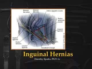
Anatomy continued: • Extends from the anterior superior iliac spine to the pubic tubercle. • About 2-3cm in length • It has an internal opening the deep ring and external opening the external ring. • It has a roof, floor, anterior and posterior wall.
Anatomy: • Roof • fibers of internal abdominal oblique and transversusabdominis muscles. • Floor • inguinal ligament throughout, with lacunar ligament added medially • Anterior wall • external abdominal oblique aponeurosis throughout, with internal abdominal oblique aponeurosis added laterally • Posterior wall • mostly transversalis fascia, with conjoint tendon (falxinguinalis), which is the joining of internal abdominal oblique and transversusabdominisaponeuroses, medially.
Openings: • deep (internal) inguinal ring • the entrance to the canal. The transversalis fascia pouches out, creating an opening through which structures can leave the abdominal cavity • superficial (external) inguinal ring • the exit from the canal. It is formed by the splitting of the diagonal fibers of the external abdominal oblique aponeurosis. Since the fibers split. A lateral crus and a medial crus are formed. The lateral crus attaches to the pubic tubercle, while the medial crus attaches to the pubic crest.
The processusvaginalis accompanies the testicle and spermatic cord structures lying against the cord in the inguinal canal.
Continued: • Contents • spermatic cord (in males) or the round ligament of the uterus (in females) as well as blood vessels, lymphatic vessels and • the ilioinguinal nerve (which enters the canal from the side, rather than passing through the deep ring).
Inguinal hernias Indirect • This enters the inguinal canal through the internal inguinal ring lateral to the inferior epigastric vessels and traverses the full length of the canal in front of the cord • They are usually congenital in origin and divided into • Bubonocoele • funicular and • complete or scrotal hernia
Direct • Usually a diffuse bulge of the medial portion of the posterior wall of the inguinal canal medial to the inferior epigastric vessels. • It is acquired Variant • Ogilvie hernia : • There is a rigid circular defect in the conjoint tendon • It is also referred to as Busoga’s hernia Direct/Indirect on the same side (Dual hernia, Pantaloon hernia or saddle bag hernia)
Examination All patients will present with a groin swelling that disappear on lying down. There may be pain or discomfort and vomiting. Inspection with the patient standing and coughing. • An indirect inguinal hernia passes downwards and medially towards the scrotum • A direct inguinal hernia protrudes directly forward in the inner part of the inguinal canal
Palpation with the patient lying down: • An indirect inguinal hernia when reducible, returns in an upward and lateral direction and is prevented from returning by pressure over the internal ring at the mid-inguinal point • A small inguinal hernia (bubonocoele) may not be detectable unless the little finger invaginates the scrotum and is passed into the external when an impulse will be felt when the patient coughs b. A direct inguinal hernia is seldom large to enter the scrotum and, when it is reducible, it returns directly backwards
Since it lies medial to the internal ring it cannot be controlled by pressure over this site and with a finger in the external ring the cough impulse is directed forward • Whether the hernia is indirect or direct it is important to assess the nature of the contents of the sac; intestine gurgles on reduction of the hernia but omentum does not • When making a local assessment of an inguinal hernia, it is important to remember the following points: • It is sometimes impossible to decide clinically whether an inguinal hernia is direct or indirect
A tense, tender, irreducible (most often an indirect hernia), in the absence of abdominal pain is simply irreducible • However, when persistent pain, loss of cough impulse and perhaps oedema and reddening of the skin over the hernia are present, together with other signs of intestinal obstruction, strangulation of the bowel must be suspected • An obstructed hernia cannot be easily distinguished clinically from a strangulated one, the distinction can only be made at operation.
Differential diagnosis • Hydrocele • Cyst of canal of Nuck • Saphenous varix • Psoas abscess • Haematoma • ascites • Maldescended testis • Femoral artery aneurysm • Pseudoaneurysm • Lymph node • Sebaceous cyst
Treatment • Herniotomy • Hernioraphy : Could be open or laparoscopic • Open • Tissue repair • Modified Bassini • Shouldice • Mcvay cooper’s ligament repair • Nylon darning • Mesh repair • Lichenstein • Plug patch • Kugel patch • Laparoscopic • Transabdominalpriperitoneal mesh repair (TAPP) • Totally extraperitonealapproah (TEP)
Femoral Hernia • A femoral hernia descends through the femoral canal beneath the inguinal ligament Variants – • Prevascular hernia (Narath’s) • This hernia passes in front of the femoral vessels. • Pectineal hernia (cloquet’s) • The hernia passes behind the femoral vessels between the pectineus muscle and the fascia • External femoral hernia (Hesselbach’s) • This passes lateral to the femoral vessels. • Langiers • The hernia passes through a defect in the lacunar ligament
Patients with femoral hernia present in one of two ways: • as a lump: • This is usually a small globular swelling situated below and lateral to the pubic tubercle • It is apparent on standing or straining but may disappear on lying down • With obstruction or strangulation: • The lump becomes tense, tender and irreducible and the overlying skin may be oedematous when strangulation is present. • In addition, the features of small bowel obstruction are apparent with abdominal pain and vomiting
Umbilical hernia Exomphalus • This is a rare neonatal condition due to an anomaly of the second stage of gut rotation when the midgut loop fails to return into the abdominal cavity during the tenth week of foetal life and present at birth as two types: i. Exomphalus Minor • The sac is small <5cm, and the umbilical cord is attached at its summit.
ii.Exomphalus Major • The sac is large >5cm and contains small and large bowel and often part of the liver. • The umbilical cord is at the inferior margin of the sac UMBILICAL HERNIA OF INFANCY • This occurs through a defect in the umbilical cicatrix during the first few days of life. • A hernial sac protrudes as a small knob at the umbilicus and is most apparent when the child cries or strains.
PARAUMBILICAL HERNIA • Appears as a large swelling to the side of the umbilical scar with features of hernia INCISIONAL HERNIA • A scar overlies the hernial swelling • It can also obstruct and strangulate
Spigelian hernia • Occurs at the level of the arcuate line • Fundus of the sac may lie beneath the internal oblique where it is impalpable • More often it may advance to spread out like a mushroom between external & internal obliques to be evident • Typically a soft reducible swelling is seen lateral to the rectus muscle, below the umbilicus • Because of rigid fascia around neck, it may strangulate
Complications of hernia • Ireducibility • Obstructed • Stangulated • Gangreneous
Conclusion • Treatment of a hernia depends on whether it is reducible or irreducible and possibly strangulated. • Reducible • Can be treated with surgery but does not have to be. • Irreducible • All acutely irreducible hernias need emergency treatment because of the risk of strangulation. • An attempt to push the hernia back can be made • Strangulation • Operation