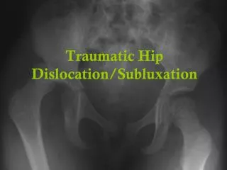
Dislocation of the hip joint
160 likes | 1.93k Vues
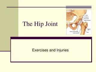
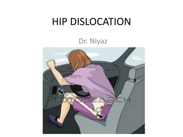
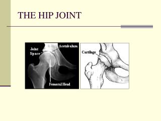
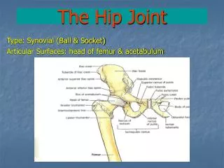
Dislocation of the hip joint . Three types of hip dislocation : - 1 . Anterior dislocation ( 10 – 15 %) 2 . Posterior dislocation ( 70 % ) 3 . Central dislocation ( rest ). Hip joint. Posterior dislocation of hip. Common in the hip joint ( 70 % ) Mechanism of injury :

Dislocation of the hip joint
E N D
Presentation Transcript
Dislocation of the hip joint Three types of hip dislocation : - 1 . Anterior dislocation ( 10 – 15 %) 2 . Posterior dislocation ( 70 % ) 3 . Central dislocation ( rest )
Posterior dislocation of hip • Common in the hip joint ( 70 % ) Mechanism of injury : • Dashbroad injury as in RTA • Simple dislocation : - Flexed knee + neutral adduction • Fracture dislocation : - flexed knee + slight abduction
Mechanism that causes the majority of dislocations is impingement (to strike )of the neck of the femoral component against the rim of the cup.
Pipkin Fracture • I - Posterior dislocation of the hip with fracture of the femoral head caudad to the fovea centralis • II - Posterior dislocation of the hip with fracture of the femoral head cephalad to the fovea centralis • III - Type I and type II with associated fracture of the femoral neck • IV - Type I, II, or III with associated fracture of the acetabulum
Clinical features • Limb shortening • Flexion , adduction and medial rotation deformity of the affected limb • Thigh rest on the contralateral limb • Head felt in the gluteal region • Movement of hip decrease • Feature of sciatic nerve palsy
Feature of sciatic nerve palsy • SCIATICA or pain localized to the hip, • PARESIS or PARALYSIS of posterior thigh muscles and muscles innervated by the peroneal and tibial nerves, • sensory loss involving the lateral and posterior thigh, posterior and lateral leg, and sole of the foot. • Pain when sitting, sneezing or coughing • tingling sensation or numbness down the leg • Foot drop
Radiology • X – ray AP and Lateral view of the pelvis showing both the hip joints • CT scan and MRI ( for acetabular fracture)
Treatment • Closed reduction ( to reduce pain ) : - • Four methods of closed reduction : - 1 . Stimson`s method : - • Position : prone , at the edge of the table • An assistant stabilizes the pelvis • Physician applies downward pressure on the calf with one hand while applying external rotation to the femur.
2 .Allis traction • Position : supine • An assistant stabilizes the pelvis • The physician simultaneously distract (to pull away ) the femur and rocks it medial to lateral . 3 . Bigelow`s method: - • Position : supine • Physician applied upwards traction on the femur while an assistant stabilize the pelvis 4 .Classical watson`s – jones method : - • Position : supine • Limb is brought to the neutral position first then longitudinal traction in the of femur is given.
After treatment • After reduction , the patient is put on a skin traction or immobilised in a Thomas split for 3 weeks . • Full weight bearing after 6 weeks . • Indication of open reduction : - • 1 . Failure of close reduction : due to obstruction by bony fragments or by soft tissues . • 2 . Instability after reduction • 3 . Sciatic nerve palsy
Complications Early : - • Sciatic nerve palsy • Irreducible fracture dislocation • Missed knee injuries • Recurrent dislocation Late : - • Myositis ossificans • Avascular necrosis of bone • Post – traumatic arthritis • Unreduced posterior dislocation