Download
1 / 50
570 likes | 1.7k Vues
SALIVARY GLAND DISEASES. omr. Introduction. Classification of salivary glands:. According to size. Major :- Parotid / Submandibular /Sublingual Minor :- Lingual / buccal / labial / palatine / glossopalatine According to nature of secretion. Serous :- Parotid Mucous :- Sublingual / minor

E N D
Classification of salivary glands: • According to size. • Major :- Parotid / Submandibular /Sublingual • Minor :- Lingual / buccal / labial / palatine / glossopalatine • According to nature of secretion. • Serous :- Parotid • Mucous :- Sublingual / minor • Mixed :- Submandibular / minor • According to location of the opening of duct. • Into vestibule :- Parotid / minor • Into oral cavity proper :- Submandibular / Sublingual / minor
Embryology • 6th-8th Weeks of Gestation • Parotid • First to develop • Last to become encapsulated • Autonomic Nervous System Crucial
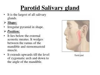
Anatomy: Parotid Gland • Wedge shaped with 5 processes • 3 Superficial • 2 Deep • Parotid Compartment • Superior – Zygoma • Posterior – EAC • Inferior – Styloid, ICA, Jugular Veins
Anatomy: Parotid Gland • 80% overlies Masseter & Mandible • 20% Retromandibular • Stylomandibular Tunnel, Isthmus of Parotid • Tail of Parotid
Anatomy: Parotid Gland • Parapharyngeal Space • Prestyloid Compartment • Poststyloid Compartment (Paragangliomas)
Anatomy: Parotid Gland • Stensen’s Duct • Arises from anterior border • 1.5 cm inferior to Zygomatic arch • Pierces Buccinator at 2nd Molar • 4-6 cm in length • 5 mm in diameter
Anatomy: Parotid Gland • Parotid Capsule • Superficial layer Deep Cervical Fascia • Superficial layer • Deep layer
Anatomy: Parotid Gland • CN VII • 2 Surgical zones • 3 Motor branches immediately • Pes Anserinus – 1.3 cm • Temperofacial Division • Cervicofacial Division • 5 Terminal branches
Anatomy: Parotid Gland • Localization of CN VII • Tragal pointer • Tympanomastoid suture • Posterior belly Digastric • Styloid process • Retrograde dissection • Mastoidectomy
Anatomy: Parotid Gland • Great Auricular nerve • Auriculotemporal nerve • Superficial Temporal vessels • Frey’s Syndrome
Anatomy: Parotid Gland • Neural compartment • VII, Great Auricular, Auriculotemporal • Venous compartment • Retromandibular vein • Arterial compartment • Superficial Temporal/Transverse Facial
Anatomy: Parotid Gland • Lymphatics • Paraparotid & Intraparotid nodes • Superficial & Deep Cervical nodes
Anatomy: Submandibular Gland • The ‘Submaxilla’ • Submandibular Triangle • Mylohyoid ‘C’ • Marginal Mandibular branch • Capsule from superficial layer of Deep Cervical fascia
Anatomy: Submandibular Gland • Wharton’s duct • Exits medial surface • Between Mylohyoid & Hyoglossus • 5 cm in length • Lingual nerve & CN XII
Anatomy: Submandibular Gland • Innervation • Superior Cervical Ganglion (symp) • Submandibular Ganglion (para) • Artery: Submental branch of Facial a. • Vein: Anterior Facial vn. • Lymphatics: Deep Cervical and Jugular chains • Facial artery nodes
Anatomy: Sublingual Gland • Between Mandible & Genioglossus • No capsule • Ducts of Rivinus +/- Bartholin’s duct • Sialogram not possible • Innervation: Same as Submandibular • Artery/Vein: Sublingual branch of Lingual & Submental branch of Facial • Lymphatics: Submandibular nodes
Anatomy: Minor Salivary Glands • 600-1,000 • Simple ducts • Buccal, Labial, Palatal, Lingual • Tumor sites: Palate, upper lip, cheek • Lingual & Palatine nn.
INVESTIGATIONS: • IMAGING • Radiography • Sialography • CT scan • MRI • Ultrasound Contd..
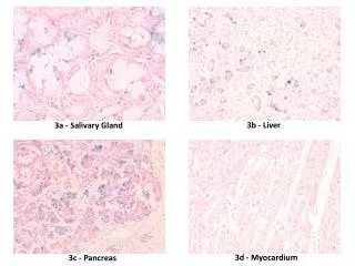
2. PATHOLOGY • Aspiration cytology & biopsy for definitive diagnosis • Frozen section • Histochemistry • Immunohistochemistry • Electron microscophy Contd..
3. FUNCTIONAL TESTS • Flow rates & sialochemistry 4. TESTS FOR RELATED / CONTRIBUTORY SYSTEMIC DISEASE • Bacteriology • Haematology • Autoantibody studies Contd..
Imaging • CT – Inflammatory • MR – Tumor • Children: U/S & MR • NO sialogram during active infection • Parotid is fatty
Microanatomy • The Secretory Unit • Acinus (serous, mucous, mixed) • Myoepithelial cells • Intercalated duct • Striated duct • Excretory duct
Microanatomy • Striated & Intercalated ducts well developed in serous, NOT mucous glands • Striated duct: HCO3 into, Cl from lumen • Intercalated duct: K into lumen, Na from lumen, producing hypotonic fluid • Excretory ducts do NOT modify saliva
Microanatomy • The Bicellular Theory • Intercalated duct • Excretory duct • The Multicellular Theory
Microanatomy • Parotid: serous & fatty • Submandibular: mixed serous • Sublingual: mixed mucous • Stroma: Plasma cells
Saliva • 600 to 700 ml (upto 1.5 L) per 24 hours. • PH is 6.7 (6.2 – 7.6) • Contribution of various glands: • Parotid:- 60 – 65% • Submandibular:- 20 – 30% • Sublingual:- 2 – 5% • Minor glands:- 6 – 7%
Composition of saliva: • Water :- 94.0 – 99.5% • Solids :- 6.0% (unstimulated), 0.5% (stimulated) • organic constituents:-urea, uric acid, glucose, aminoacid, lactate, fatty acids, proteins like amylase, peroxidase, lysosyme, IgA, IgM, IgG. • inorganic constituents:- Ca, Mg, F, HCO3, K, Na, Cl, NH4. • gases:- CO2, N2, O2. • constituents from oral cavity:- desquamative epithelial cells, bacteria.
Digestion (Amylase, Lipase) Antibacterial (Lysozyme, IgA, Peroxidase, FLOW) Mineralization Protective Pellicle Inhibition of dental caries Water balance Lubrication action Taste of food Buffering action Hygienic action Function of Saliva
Factors influencing salivary secretion: • Taste and smell • Mechanical stimulation of oral mucosa and gingiva • Mastication of food • Chemical irritation of oral mucosa & stomach • Distension / irritation of oesophagus • Pregnancy
Causes of salivary gland dysfunction: • Drugs (anticholinergic, antihistamine, antihypertensive) • Radiation theraphy • Oncologic chemotheraphy • Psychological factors • Systemic diseases • Malnutrition and neoplasms
Clinical features: • Commonly present as an asymptomatic mass • Pain – infection/ hemorrhage/ infected cyst • Malignant lesions are also symtomless unless involves the nerves • Nasal obstruction – tumor involves paranasal sinuses / nose • Xerostomia
Salivary hypofunction • Candidiasis • Lichen Planus • Burning Mouth • Aphthous ulcers • Dental caries • Xerostomia not reliable
XEROSTOMIA • Causes; • Depression / chronic anxiety • Dehydration • Drugs • Diseases
XEROSTOMIA • Clinical features; • Dry mouth • Salivary froath adhering to mucosa • Rapid dental decay • Adherence of food debris to teeth • Soreness / halitosis
XEROSTOMIA • MANAGEMENT • Preventive theraphy • Symptomatic treatment • Local or topical salivary stimulation • Systemic salivary stimulation
Effects of Aging • Total salivary flow independent of age • Acinar cells degenerate with age • Submandibular gland more sensitive to metabolic/physiologic change • Unstimulated salivary flow more greatly affected by physiologic changes
DEVELOPMENTAL APLASIA / HYPOPLASIA ACCESSORY SALIVARY DUCTS DIVERTICULI DARIER’S DISEASE ACQUIRED REACTIVE LESIONS INFECTIONS METABOLIC CONDITIONS ASSOCIATED IMMUNE DEFECTS NEOPLASMS CLASSIFICATION OF DISEASES
BENIGN Pleomorphic adenoma Monomorphic adenoma Ductal papilloma MALIGNANT Mucoepidermoid carcinoma Adenoid cystic carcinoma Acinic cell carcinoma Salivary duct carcinoma Squamus cell carcinoma Basal cell adenocarcinoma Adenocarcinoma NEOPLASMS