Elbow Trauma
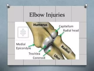
Elbow Trauma. Elbow Trauma. 6% of all fractures and dislocations involve elbow Most common fractures differ between adults and children M.C. in adults- radial head and neck fxs. M.C. in children- supracondylar fxs. Complex anatomy requires 4 views for adequate interpretation

Elbow Trauma
E N D
Presentation Transcript
Elbow Trauma • 6% of all fractures and dislocations involve elbow • Most common fractures differ between adults and children • M.C. in adults- radial head and neck fxs. • M.C. in children- supracondylar fxs. • Complex anatomy requires 4 views for adequate interpretation • AP in extension, medial oblique, lateral and axial olecranon (Jones view)
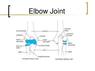
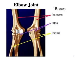
Normal Elbow Anatomy • Very important to be aware of pediatric growth centers • CRITOE http://www.radiologyassistant.nl/en/4214416a75d87 http://med_practice.byethost7.com/wp2/?p=21
Normal Alignment • Anterior humeral line- line drawn along anterior surface of humeral cortex should pass through the middle third of the capitellum • Radiocapitellar line- Line drawn through the proximal radial shaft and neck should pass through to the articulating capitellum http://imageinterpretation.co.uk/elbow.html
Signs of Fracture • Usual signs may not be readily visible • Fracture line, cortical disruption, etc. • Soft tissue signs can indicate fracture • Fat pad sign • On lateral, might see fat pad parallel to anterior humeral cortex, but should never see posterior fat pad • With effusion, anterior may be displaced and will be shaped like a sail (sail sign)
Fat Pad Sign • Posterior fat pad is normally buried in olecranon fossa and not visible • Becomes elevated and visible with joint uffusion • Effusion (acute capsular swelling) can be from any origin (hemorrhagic, inflammatory, infectious, traumatic, etc.) • Ant. fat pad may be obliterated, so post. Fat pad is more reliable when visible
Distal humerus fractures • 95% extend to articular surface • Classified according to relationship with condyle and shape of fracture line • Supracondylar, intercondylar, condylar and epicondylar
Supracondylar Fractures • Most common elbow fracture in children (60%) • Fracture line extends transversely or obliquely through distal humerus above the condyles • Distal fragment usually displaces posteriorly Normal
Intercondylar fracture • Fracture line extends between medial and lateral condyles and extends to supracondylar region • Results and T or Y shaped configuration for fracture • Called trans-condylar if it extends through both condyles
Epicondylar fracture • Usually avulsion from traction of respective common flexor (medial) or extensor (lateral) tendons • Medial epicondyle avulsion common in sports with strong throwing motion (little leaguer’s elbow)
Fractures of Proximal Ulna • Olecranon fx.- direct trauma or avulsion by triceps tendon • Coronoid process fx.- avulsion by brachialis or impaction into trochlear fossa • Rarely isolated; usually associated with post. elbow dislocation
Fractures of Proximal Radius • M.C. adult elbow fx. (50%) • FOOSH transmits force causing impaction of radial head into capitellum • Chisel fracture- incomplete fracture of radial head that extends to center of articular surface • Usual rad. signs (fx. Line, articular disruption) may not be visible • May be occult; fat pad sign is good indicator of occult fx.
Fractures of the forearm • Isolated ulnar fractures • Isolated radial fractures • Bony rings usually can't be fractured in one place without disruption somewhere else in the ring • 60% or forearm fractures involve both bones (BB fractures) • These fractures usually have associated displacement with angulation and rotation
Isolated Ulnar Fractures • Distal shaft (Nightstick fx.)- direct trauma • Proximal shaft (Monteggia’s fx.)- fx. of proximal ulna with dislocation of radius http://radiographics.rsna.org/content/24/4/1009/F31.expansion.html http://www.wheelessonline.com/ortho/monteggias_fracture
Isolated Radial Fractures • Most frequent is a Galeazzi’s fx. (reverse Monteggia’s fx.) • Fracture of distal radial shaft with dislocation of distal radioulnar joint • Rare, but serious injury http://www.learningradiology.com/archives05/COW%20157-Galeazzi%20Fx/galeazzicorrect.htm
Dislocations of Elbow • 3rd m.c. dislocation in adults behind shoulder and interphalangeal joints • More common in children • Classified according to displacement of radius an ulna relative to humerus • Posterior, posterolateral, anterior, medial and anteromedial • Posterior and posterolateral or more most common • 85-90% of all elbow locations • 50% have associated fractures
Pulled Elbow • AKA nursemaid’s elbow • Occurs when child’s hand is pulled, tractioning arm and causing radial head to slip out from under annular ligament and trapping the ligament in the radiohumeral articulation • Immediate pain; stuck in mid-pronation due to pain • No radiographic pain • Supination reduces the dislocation and ends pain, usually during positioning of lateral radiograph
References Yochum, T.R. (2005) Yochum and Rowe’s Essentials of Skeletal Radiology, Third Edition. Lippincott, Williams and Wilkins: Baltimore.