Download

1 / 26
310 likes | 1.89k Vues
Uterine Leiomyoma. UNC School of Medicine Obstetrics and Gynecology Clerkship Case Based Seminar Series. Objectives for Uterine Leiomyoma . Discuss the prevalence of uterine leiomyomas Describe the symptoms and physical findings in patients with uterine leiomyomas

E N D
Uterine Leiomyoma UNC School of Medicine Obstetrics and Gynecology Clerkship Case Based Seminar Series
Objectives for Uterine Leiomyoma • Discuss the prevalence of uterine leiomyomas • Describe the symptoms and physical findings in patients with uterine leiomyomas • Describe the diagnostic methods to confirm uterine leiomyomas • List the management options for the treatment of uterine leiomyomas
Patient presentation • A 42-year-old G3 P3 female presents with a history of abnormal bleeding and pelvic pain. She was well until approximately age 35, when she began developing dysmenorrhea and progressive menorrhagia. The dysmenorrhea was not fully relieved by NSAIDS. Over the next several years, the dysmenorrhea and menorrhagia became more severe. She then developed intermenstrual bleeding and spotting, as well as pelvic pain, which she describes as a constant feeling of pressure. She also complains of urinary frequency.
Patient presentation • Past gynecological history is otherwise non-contributory. She delivered three children by caesarean section, the last with a tubal ligation at age 30. Her past medical history is unremarkable.
Physical Exam • Reveals a well-developed, well-nourished woman in no distress. Vital signs and general physical exam are unremarkable. Abdominal examination reveals an irregular-sized mass into extending halfway between the pubic symphysis and umbilicus and to the right of the midline. Pelvic exam reveals a normal appearing vagina and cervix. The uterus is markedly enlarged and irregular, especially on the right side where it appears to reach the lateral pelvic sidewalls. The examiner is unable to palpate normal ovaries due to the mass.
Patient PresentationDiagnostic Evaluation • Laboratory • Beta HCG is negative. CBC reveals hemoglobin of 10.3 and hematocrit of 31.2. Indices are hypochromic, microcytic. Serum ferritin confirms mild iron deficiency anemia. Pap smear is normal with no evidence of dysplasia. Endometrial biopsy reveals proliferative endometrium. ECC is negative for malignancy. Ultrasound shows a large irregular mass, filling the pelvis and extending into the lower abdomen. The mass does extend into the right side of the pelvis. The ovaries are not visualized.
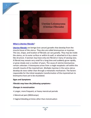
Definition • Uterine leiomyomas (“fibroids”) are benign tumors derived from the smooth muscle cells of the myometrium
Prevalence • Approximately 45% of women have uterine leiomyomas by the 5th decade of life • Vast majority are asymptomatic • Primary indication for 200,000 hysterectomies in the U.S. each year • Sarcomatous changes occur in < 0.1%
Risk Factors • Increasing age during reproductive years • Ethnicity (African American) • Nulliparity • Family history
Pathogenesis of Leiomyomas • Factors that initiate leiomyomas unknown • Estrogen and progesterone important to growth • Increased levels of estrogen and progesterone receptors present • Estrogen induces proliferation of smooth muscle cells • Progesterone produces proteins which prohibit apoptosis • Increased levels of growth factors produce fibronectin and collagen
Characteristics of Leiomyomas • Spherical, well-circumscribed, white, firm lesions • Always arise within the myometrium (intramural) • Migrate to various anatomic locations • Submucosal – toward endometrium • Intramural – within myometrium • Subserosal – toward serosal surface • Pedunculated and/or parasitic • Poor internal blood and lymphatic supply • Cystic degeneration • Calcification
Anatomic Locations Pedunculatedsubserosal Uterus Pedunculated submucosal Subserosal Intramural Submucosal Vagina
Clinical Manifestations Clinical Manifestations Bleeding symptoms • Menorrhagia – heavy bleeding • Metrorrhagia – bleeding between menses • Dysmenorrhea – painful menses Bulk symptoms • Pelvic pressure • Urinary frequency • Infertility and/or recurrent pregnancy loss *Many women are asymptomatic; symptoms depend on size and location of fibroids
Physical Exam • Abdominal exam • Palpable mass if uterus > 12-14 wk gestational size • Pelvic exam • Firm, irregularly enlarged uterus • Midline, occasionally adnexal • Mass displaced with cervix • Usually nontender
Differential Diagnosis • Uterine sarcoma • Ovarian neoplasm • Tubo-ovarian inflammatory mass • Diverticular/inflammatory bowel mass • Colon cancer • Pelvic kidney
Diagnosis • Bimanual pelvic exam • Transvaginal ultrasound (TVUS) • Sonohysterography • Hysterosalpingography • Diagnostic hysteroscopy • MRI
Pathology Well circumscribed white tan firm masses with a whorled appearance
Pathology Microscopically leiomyomas are composed of bland smooth muscle. They can be more fibrotic than this example or more cellular.
Patient presentations • 42yo P2 s/p BTL with 16 week size uterus, menorrhagia, anemia, bulk symptoms Management options? • 32yo G0 who desires fertility with otherwise the same presentation? Management options? • 42yo P3 s/p BTL with bleeding sx, no bulk sx and a normal size uterus Could she still have fibroids? Management options?
Management (Surgical) *Intervention for patients with leiomyomas not amenable to medial therapy
Management (Surgical) Desire future fertility… • Myomectomy • Laparotomy – larger fibroids • Laparoscopic – pedunculated or subserosal fibroids • Hysteroscopic – submucosal fibroids, >50% in cavity Desire uterine preservation but not fertility… • Endometrial ablation • Uterine artery emboloization (UAE) No desire for uterine preservation or fertility… • Hysterectomy (definitive) • Laparotomy (TAH) – larger fibroids • Laparascopic (TVH, TLH) – smaller fibroids
Patient presentations • 34 yo P1 with menorrhagia and dysmenorrhea with an 8-10 weeks size uterus Management options? • What is this same patient were asymptomatic?
Management (Medical) 1st line treatment • NSAIDS • Progestin-only therapies (Depo Provera, Mirena IUD) • Combination therapies (OCP’s, patches, vaginal rings) 2nd line treatment • GnRH analog (Lupron) – blocks endometrial proliferation, shrinks myometrium, and reduces leiomyoma volume • Causes vasomotor symptoms (hot flashes) and bone loss • Short courses, used primarily for pre-surgical shrinkage of leiomyoma • GnRH analog + hormonal agents • Minimize adverse hypoestrogenism effects • Mifepristone (RU 486) – progesterone receptor antagonist • Still experimental, shown to reduce volume by 50% over 3 months
Management (Conservative) • Treatment is not necessary if…. • Asymptomatic • Fibroid small (<12 wk gestational size) • Near menopause
Bottom Line Concepts • Most uterine leiomyomas are symptomatic and require no intervention. • Uterine leiomyomas can cause excessive uterine bleeding, pelvic pressure and pain, and infertility. • Fibroids can be subserosal, intramural, or submucosal. Prolonged or heavy bleeding may be associated with intramural or submucosalmyomas. • Conservative or medical management should be considered prior to surgical management. • Treatment options for leiomyoma include myomectomy, endometrial ablation, uterine artery embolization and hysterectomy. • Pregnancies in women with fibroids are usually uneventful. • Fibroids are rarely the cause of infertility. In women who have a myomectomy in which the endometrial cavity is entered, future deliveries must be by cesarean birth.
References and Resources • APGO Medical Student Educational Objectives, 9th edition, (2009), Educational Topic 53 (p114-115). • Beckman & Ling: Obstetrics and Gynecology, 6th edition, (2010), Charles RB Beckmann, Frank W Ling, Barabara M Barzansky, William NP Herbert, Douglas W Laube, Roger P Smith. Chapter 44 (p389-392). • Hacker & Moore: Hacker and Moore's Essentials of Obstetrics and Gynecology, 5th edition (2009), Neville F Hacker, Joseph C Gambone, Calvin J Hobel. Chapter 19 (p241-245).