3. Methods
1. Purpose. 2. Conclusions. 3. Methods. Increased patient throughput for treatment with helical tomotherapy. K. Petersson, 1 C. Ceberg, 1 T Knöös, 1,2 and M. Enmark 2 1 Medical Radiation Physics, Lund University, Lund, Sweden 2 Radiation Physics, Skåne University Hospital, Lund, Sweden.

3. Methods
E N D
Presentation Transcript
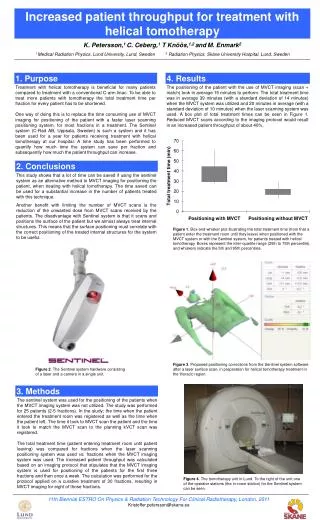
1. Purpose 2. Conclusions 3. Methods Increased patient throughput for treatment with helical tomotherapy K. Petersson,1 C. Ceberg,1 T Knöös,1,2 andM. Enmark2 1 Medical Radiation Physics, Lund University, Lund, Sweden 2 Radiation Physics, Skåne University Hospital, Lund, Sweden 4. Results Treatment with helical tomotherapy is beneficial for many patients compared to treatment with a conventional C-arm linac. To be able to treat more patients with tomotherapy the total treatment time per fraction for every patient has to be shortened. One way of doing this is to replace the time consuming use of MVCT imaging for positioning of the patient with a faster laser scanning positioning system, for most fractions in a treatment. The Sentinel system (C-Rad AB, Uppsala, Sweden) is such a system and it has been used for a year for patients receiving treatment with helical tomotherapy at our hospital. A time study has been performed to quantify how much time the system can save per fraction and subsequently how much the patient throughput can increase. The positioning of the patient with the use of MVCT imaging (scan + match) took in average 15 minutes to perform. The total treatment time was in average 39 minutes (with a standard deviation of 14 minutes) when the MVCT system was utilized and 20 minutes in average (with a standard deviation of 10 minutes) when the laser scanning system was used. A box plot of total treatment times can be seen in Figure 1. Reduced MVCT scans according to the imaging protocol would result in an increased patient throughput of about 40%. This study shows that a lot of time can be saved if using the sentinel system as an alternative method to MVCT imaging for positioning the patient, when treating with helical tomotherapy. The time saved can be used for a substantial increase in the number of patients treated with this technique. Another benefit with limiting the number of MVCT scans is the reduction of the unwanted dose from MVCT scans received by the patients. The disadvantage with Sentinel system is that it scans and positions the surface of the patient but we almost always treat internal structures. This means that the surface positioning must correlate with the correct positioning of the treated internal structures for the system to be useful. Figure 1. Box-and-whisker plot illustrating the total treatment time (from that a patient enter the treatment room until they leave) when positioned with the MVCT system or with the Sentinel system, for patients treated with helical tomotherapy. Boxes represent the inter-quartile range (25th to 75th percentile), and whiskers indicate the 5th and 95th percentiles. Figure 3. Proposed positioning corrections from the Sentinel system software after a laser surface scan, in preparation for helical tomotherapy treatment in the thoracic region. Figure 2. The Sentinel system hardware consisting of a laser and a camera in a single unit. The sentinel system was used for the positioning of the patients when the MVCT imaging system was not utilized. The study was performed for 25 patients (2-5 fractions). In the study; the time when the patient entered the treatment room was registered as well as the time when the patient left. The time it took to MVCT scan the patient and the time it took to match the MVCT scan to the planning kVCT scan was registered. The total treatment time (patient entering treatment room until patient leaving) was compared for fractions when the laser scanning positioning system was used vs. fractions when the MVCT imaging system was used. The increased patient throughput was calculated based on an imaging protocol that stipulates that the MVCT imaging system is used for positioning of the patients for the first three fractions and then once a week. The calculation was performed for the protocol applied on a curative treatment of 30 fractions, resulting in MVCT imaging for eight of those fractions. Figure 4. The tomotherapy unit in Lund. To the right of the unit one of the operator stations (the in-room station) for the Sentinel system can be seen. 11th Biennial ESTRO On Physics & Radiation Technology For Clinical Radiotherapy, London, 2011 Kristoffer.petersson@skane.se