Modeling Mitosis With Playdoh Experiment
Learn about cell division stages in mitosis through hands-on playdoh modeling. Capture your models on paper plates and enhance your understanding of PMAT phases. Engage in interactive learning with visuals and photography.

Modeling Mitosis With Playdoh Experiment
E N D
Presentation Transcript
The Cell Cycle Section 10-2 G1 phase M phase S phase G2 phase
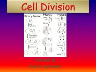
At the end of G2Mitosis begins – Mitosis is divided into 4 Phases 1. PROPHASE: • Nuclear membranebegins to disappear • DNA condenses and chromatidscome together to form Chromatid pairs(aka sister chromatids) held together by the Centromere • Centriolesappear, migrate to the cells pole and spindle fibers appear
Spindle Fibers:framework of microtubules that guide the movement of chromosomes during mitosis and meiosis.
2. METAPHASE • All the chromosomes are in a plane (line) in the middle of the cell. • Spindles are fully formed and attached to the chromosomes at the centromeres
3. ANAPHASE • In anaphase, chromatid pairs separate as the spindle fibers pull them apart and toward each pole
4. TELOPHASE • Chromatid begin to unwind and relax • Nuclear membrane reforms • Telophasecontinues until the cells split The process of cells actually splitting is called cytokinesis.
Modeling Mitosis With Playdoh Materials: Five paper plates 4 different playdoh colors Procedure: Assign a playdoh color to represent Mother’s chromosome_____________ Father’s chromosome______________ Centrioles_______________ Spindle Fibers_______________ 2. Label each plate to represent the four stages of mitosis (PMAT) and cytokinesis 3. Use the graphics in the mitosis powerpointor from chapter 9 in iBooks to model the stages of mitosis using the playdoh 4. When all five plates have completed models please use your phone (or my phone) to take a picture and upload the photo to notability. Once the photos are in notability please label the chromatids, sister chromatids (if present), centrioles, and spindle fibers.