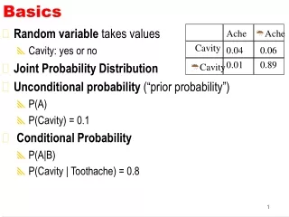

Particle Physics Basics for Science Graduates
E N D
Presentation Transcript
Basics This course is designed for persons with an undergraduate degree in science or engineering but no knowledge of how particles (particularly protons) interact with matter. (To call it ‘particle physics’ would be too grandiose.) The best texts were written around the 1950’s: Bruno Rossi, ‘High-Energy Particles,’ Prentice-Hall (1952) Emilio Segrè, ‘Experimental Nuclear Physics,’ Wiley (1953) David M. Ritson, ‘Techniques of High Energy Physics,’ Interscience (1961) They are hard to get, and of course cover tons of stuff that’s either out-of-date or not needed. While we’re at it, two excellent books much more focused on our particular topic Eric J. Hall, ‘Radiobiology for the Radiologist,’ Harper and Row (1978) M.R. Raju, ‘Heavy Particle Radiotherapy,’ Academic Press (1980) are great for background. This lecture defines fundamental quantities that we’ll need throughout the course, and explores the relation between dose rate and beam current.
Energy (MeV) The MKS unit of energy is the joule (J). In particle physics, however, we normally use the electron volt (eV) or mega electron volt (MeV). The electron volt is defined as the kinetic energy gained by one electron in falling through a potential difference of 1 volt. Since and the magnitude of the electron charge is 1.602 × 10⁻¹⁹ C it follows that or Binding energies of atomic electrons are of order eV. Energy to remove a proton or neutron from the nucleus is of order MeV. Proton energies of concern in radiotherapy are 3-300 MeV.
Areal Density (g/cm2) The range of a 160 MeV proton is 17.6 cm in water. In air it is 16700 cm (167 m). Most of this very large difference comes from the fact that air under normal conditions is a gas with a density of only 0.0012 g/cm3. To reveal the underlying physics, that is, to eliminate the essentially trivial role of density, we normally think of a degrader not in terms of its thicknessΔx (cm) but its areal density The two are the same for water, whose density ρ is 1 g/cm3 by definition. By this measure the range of our 160 MeV proton in air is 20.0 g/cm2 so we see that the stopping powers of air and water are not so different after all. There is no special symbol for areal density. In computation it is convenient to think of it as thickness × density, but if you have a degrader and wish to measure its areal density you should weigh it and divide by the area, usually a much more accurate method.
Mass Stopping Power (MeV/(g/cm2)) When a proton slows down its specific energy change dE/dx is negative: as x increases, E decreases. The positive quantity S ≡ – dE/dx (MeV/cm) is called the stopping power of the material. It depends on the material and on the proton’s speed (or energy) in a way we’ll discuss later. We rarely use stopping power by itself. Instead we use the mass stopping power The mass stopping power of 160 MeV protons in water is 5.2 MeV/(g/cm2). We’ll generally use z as the beam direction and T as the kinetic energy, so to be strictly consistent we should have denoted the stopping power –dT/dz . However, everyone calls it dE/dx . This is just one example of the inevitable inconsistency in notation when we deal with an extensive scientific literature.
Fluence (protons/cm2) Fluence characterizes the number of protons in a beam. In beam line design the protons always travel in the same direction ± a few degrees so we don’t need the general definition of fluence in terms of a sphere. Instead, we can just use a plane element of area and define fluence by where dA is an infinitesimal element of area perpendicular to the beam and dN is the number of protons passing through it. The fluence rate is sometimes denoted by lowercase φ and earlier called ‘flux’. Fluence and fluence rate are scalar fields: they are directionless quantities that may, and usually do, vary with x, y, z and t.
Physical Dose (Gy = J/kg) The physical absorbed dose (or just ‘dose’) at a point in a radiation field is the energy absorbed per unit target mass The J/kg has a special name, the gray or Gy. A typical course of treatment is (very roughly) 70 Gy, usually delivered in fractions of (roughly) 2 Gy each to the target volume. A whole-body dose of 1-2 Gy causes radiation sickness within a few hours, usually with recovery in a few weeks. Whole-body doses in the range 2-6 Gy are usually fatal within 2 months; for survivors, recovery takes up to a year (Glasstone, ‘Sourcebook on Atomic Energy,’ van Nostrand, 1967). An older unit of dose still in frequent use is the rad (‘radiation absorbed dose’): 1 rad ≡ 100 erg/g = 0.01 Gy. Radiation oncologists often hedge by using the term ‘centiGray’ (cGy) instead of ‘rad’. Dose is also a scalar field. It frequently happens that the energy lost by the radiation is greater than the energy absorbed by the target volume. We will see this in the case of protons.
Equivalent Dose, RBE (Sv) In this course we are mainly concerned with supplying a prescribed physical dose to a prescribed volume. However, dose all by itself is not a very good measure of biological effect. Its advantages are that it’s easy to define and relatively easy to measure. (In the early days a quantity called exposure, a measure of the ionization produced, was used instead.) The biological effect of a given dose depends on the type of radiation, the target tissue, the fraction of an organ exposed and the timing of dose delivery (fractionation). In radiobiology, the ‘relative biological effectiveness’ (RBE) of a radiation type is defined as the ratio of the dose of a standard radiation, frequently 60Co γ rays, to the dose of the radiation in question that gives the same biological effect. The RBE of therapy protons is generally 1-1.1 . In other words, their biological effect, dose for dose, is not greatly different from 60Co, although the very low energy protons at the distal edge of the Bragg peak may have an RBE as high as 1.65 (Coutrakon et al. (Med. Phys. 24 (1997) 1499). Equivalent dose has its own units: sieverts (Sv) correponding to Gy and rem (‘roentgen equivalent man’) corresponding to rad. The average American receives an annual dose 360 millirem = 3.6 mSv per year, about 80% from natural sources. A roundtrip transcontinental flight gets you an extra 5 mrem.
area A N protons Δx The Fundamental Formula The equation relating dose to fluence and stopping power is the starting point of most beam line design problems. From the figure : dose = fluence × mass stopping power
D = Φ S/ρ in Practical Units J/Kg = Gy is a perfectly good unit of dose but (protons/m2) for fluence and J/(Kg/m2) for mass stopping power are not convenient. A better form is where Φ is in Gp/cm2 and S/ρ is in MeV/(g/cm2). The gigaproton = 1 Gp = 109 protons is a handy unit for proton therapy. If we use charge areal density instead of fluence that gets rid of the constant and we find where q/A is the total proton charge divided by the area through which it passes (nC/cm2) (nC = nanoCoulomb). Finally, taking the time derivative where iP/A is the proton current density (nA/cm2). Therapy doses are of the order of 1 Gy, target areas are of the order of cm2, and S ≈ 5 MeV/(g/cm2) (160 MeV protons in water) so we have already learned something useful. A proton therapy accelerator needs to deliver nanoAmps to the proton nozzle.
Dose Rate in a Therapy Beam The last formula leads to a ±10% formula for the dose rate in a scattered and range-modulated proton beam. The derivation anticipates a lot of stuff we’ll cover later, but let’s try it. Our model beamline has a modulator which doubles as the first scatterer S1, a contoured second scatterer S2 and a water tank (element 3). The scattering system is designed to deliver uniform fluence to a circle of area A with an efficiency ε = 0.45 . For the peak/entrance ratio of a pristine Bragg peak we’ll write fBP which is always close to 3.5 and for the fractional time of the thinnest modulator step we’ll write fMOD.
Dose Rate in a Therapy Beam (continued) Our formula applies at the entrance to the water tank and the derivation consists basically of working both forwards and backwards from there because we want the dose rate at mid-SOBP as a function of proton current into S1. First, we need a factor ε on the RHS because the current into A is lower than the input current by that factor. Next, insert a factor fBP because the dose at the distal corner of the SOBP is higher by that factor. (The average dose rate anywhere in the SOBP is the same as the distal corner.) If there is no range modulation (S1 uniform) we are done. If there is, we need to notice that the distal layer is only irradiated for a fractional time fMOD ( = 1 for no mod, ≈ 0.35 for full mod) and insert that factor as well. Putting it all together with units nA/cm2 and MeV/(g/cm2) as before. iP is assumed constant and dose rate is averaged over 1 modulator cycle. The next slide shows fMOD as a function of relative modulation.
fMOD vs. Fractional Modulation fMOD depends mainly on the shape of the Bragg peak and relatively little on details of the scattering system. The main point here is that, although dose per proton varies exactly as the inverse area of the design field, its dependence on modulation (the longitudinal extent of the field) is more complicated. However, it is always lowered by roughly a factor 3 as we go from no mod to full mod. Or, it takes about 3 times as many protons to treat full mod to a given dose.
The Gaussian Function The Gaussian is an extremely important function having (in the most general case) 3 parameters. It, and its first three moments, are given below. The next slide is a graphical illustration. (The parameters are usually not called a, b, c .)
The Gaussian and its Parameters(graphical) ymax 0.606× ymax c area a b
The Error Function Often, the dose falloff at the edge of a field is described by the error function (see following slide and H.B. Dwight, ‘Tables of Integrals and Other Mathematical Data,’ Macmillan (1947)) : where The integral of a standard Gaussian between arbitrary limits is To find the 80% point let x1 = - ∞ , x2 = x80 and the integral be 0.8 : → Treating the 20% point similarly we find the 80/20 penumbra
The Error Function (continued) So if the rms value y0 of the transverse variable (here called y) happens to be 1 mm, the 80/20% penumbra width is 1.68 mm. Finding y0 is the real problem, which will be addressed later.
Relativistic Proton Kinematics Therapy protons are relativistic: the beam at HCL (160 MeV) traveled at just about half the speed of light. Fortunately, you can relate anything to anything else for a proton by juggling just three formulas. They are in which c = speed of light, v = proton speed, p = proton momentum, T = proton kinetic energy, E = proton total energy and mc2 = proton rest mass = 938.27 MeV. For instance, you should be able to show that the quantity pv, used in multiple scattering theory, is given by or that 160 MeV protons have β = 0.5 as claimed. Hint: don’t separate the c from pc or mc2 (leave every term in units of energy as long as possible).
Sample Derivation Here’s a derivation of the result given in the previous slide: This particular form of the result is handy because the nonrelativistic (small T) and relativistic (large T) limits are obvious: 2T and T respectively. The former limit also agrees with the familiar T = mv2/2.
Three Handy Formulas In proton radiotherapy we most frequently think in terms of the proton kinetic energy T (MeV) and we want to express other kinematic quantities in terms of T. The three equations below cover most requirements. τ is the reduced kinetic energy τ ≡ T/mpc2 where mpc2 is the proton rest energy, 938.27 MeV. The second is, of course, the one we just proved. You should be able to prove the other two. All three have obvious relativistic and non-relativistic limits, and avoid calculating the difference between two large quantities as sometimes happens in relativistic math.
Summary We have defined basic quantities we’ll need throughout the course: eV or MeV unit of energy measurement; areal density (g/cm2) ; mass stopping power MeV/(g/cm2) ; fluence (protons/cm2) and dose (gray = Gy = joule/Kg). We have derived the fundamental formula relating dose to fluence and mass stopping power which, in practical units, reads with the RHS in nC/cm2 and MeV/(g/cm2), and from that derived a practical formula for the dose rate in a therapy beam, given the incident proton current. WARNING Our formula, or for that matter the output of any beam line design program, should never be used to determine the dose to a patient, but only to estimate the current required for a given dose rate etc. etc. Dose to the patient must be determined either with a calibrated dosimeter in a simulated treatment field or with a validated computer model of the beam monitor output factor (‘model calibration’).