ENZYMES
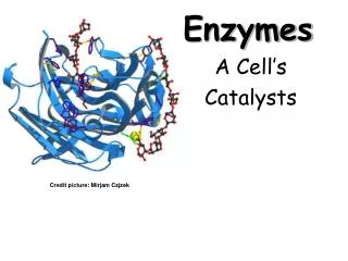
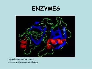
ENZYMES. Crystal structure of trypsin http://en.wikipedia.org/wiki/Trypsin. Function of enzymes. ● they are special proteins produced by living cells they are catalysts → increase the rate of chemical reactions and decrease the activation energy of the reaction

ENZYMES
E N D
Presentation Transcript
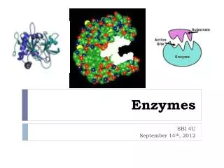
ENZYMES Crystal structure of trypsin http://en.wikipedia.org/wiki/Trypsin
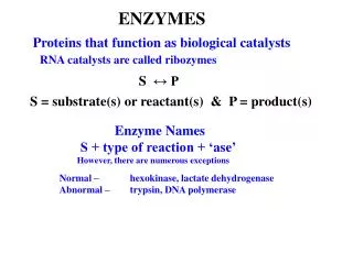
Function of enzymes ●they are special proteins produced by living cells • they are catalysts→ increase the rate of chemical reactions and decrease the activation energy of the reaction • the action of most enzymes is very specific – substrate and reaction specifity
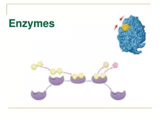
Enzyme catalysis Enzymes E are able to specifically bind the reactants (their substrates S) at the active site → complex E-S (transition state, ↓ activation energy) → destruction of complex E-S to products Pand E http://en.wikipedia.org/wiki/Enzyme
Characteristics of enzymes • Intracellular enzymes • Extracellular enzymes • Simple enzymes – only protein structure • Complex enzymes = protein structure + cofactor Cofactors are nonprotein compounds. Cofactor can be: 1) inorganic element: Zn2+, Mn2+, Mg2+, Fe2+, Cu2+, …. 2) organic molecule a) coenzymes are slightly bound to the enzyme, undergo a chemical change and are released: NAD(P)+, FAD, coenzyme Q,.. b) prosthetic groups are tightly bound to the enzyme and remain associated with enzyme during reaction: heme, …
Coenzymes NAD+↔ NADH + H+FAD↔ FADH2 nicotinamide adenine dinucleotide flavin adenine dinucleotide (vit. B2 = riboflavin) Other examples: coenzyme A, coenzyme Q, tetrahydrofolate, thiamine diphosphate (vit. B1 = thiamine) http://web.indstate.edu/thcme/mwking/vitamins.html
Prosthetic groups Biotin (vit. H) Heme Another example: pyridoxal phosphate (derivate of vitamine B6) http://web.indstate.edu/thcme/mwking/vitamins.html
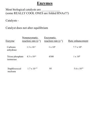
Nomenclature of enzymes 1) The first discovered enzymes were named according to their source: name of enzyme + suffix-in Pepsin is found in the gastric juice (Greek pepsis = digestion). 2) Enzymes were named according to their substrate: name of substrate + suffix –ase Lipase catalyzes the hydrolysis of lipids. Urease catalyzes the hydrolysis of urea. 3) In 1961 International Union of Biochemistry recommended that enzymes be systematically classified according to the general type of reaction they catalyze → 6 major classes. Each enzyme has a EC number (four-digit number) Lactate dehydrogenase has the EC number 1.1.1.27
Classification of enzymes • Oxidoreductasescatalyze redox reactions alcohol dehydrogenase oxidases, oxygenases, peroxidases, catalase 2. Transferasescatalyze the transfer of functional groups between donors and acceptors aminotransferases, kinases 3. Hydrolasescatalyze the hydrolytic cleavage of substrates peptidases, proteases, lipases, α-amylase 4. Lyases (synthases)catalyze non-hydrolytic and non-oxidation cleavage or synthesis of molecules(removing/addition of the small molecule from/to substrate) carboxylases/decarboxylases, hydratases/dehydratases 5. Isomerasescatalyze intramolecular changes in substrate molecules epimerases, mutases 6. Ligases (synthetases)catalyze synthetic reactions where 2 molecules are joined at 1 molecule, synthesis requires an energy (ATP) polymerases
Enzyme kinetics The Michaelis-Menten model Michaelis constant KM corresponds to the substrate concentration [S] at which velocity V is half of the maximum velocity Vmax (when v = ½ Vmax). An enzyme with a high affinity for its substrate has a low KM value. KM = mol/L http://en.wikipedia.org/wiki/Enzyme
Enzyme kinetics • Lineweaver-Burk plot provides a useful graphical method for analysis of the Michaelis-Menten equation: • taking the reciprocal gives Figure was found at http://en.wikipedia.org/wiki/Lineweaver-Burk_diagram
Enzymatic activity Reaction rate is expressed as a change in concentration per unit time (mol/L / s). For enzyme-catalyzed reaction: substrate turnover per unit time is commonly used: ● Unit:katal (kat) = mol of substrate / s kat and nkat are used in medicine ● International unit:IU = μmol of substrate / min 1 kat = 6 x 10⁷ U
Factors that influence enzyme activity • Concentration of substate The rate of an enzymatic reaction increases as the substrate concentration increases until a limiting rate is reached. • Concentration of enzyme Enzyme concentration is much lower than the concentration of substrate. The rate of an enzyme-catalyzed reaction is directly dependent upon the enzyme concentration.
●Temperature Most enzymes of warm-blooded animals have temperatures optimum of about 37 oC. Protein structure of enzymes is denatured by heat (above 55 oC) • Hydrogen ion concentration (pH) Extreme values of pH (low/high) cause denaturation of protein. Optimum pH of enzyme is a narrow pH range. Optimal pH for pepsin is 2.0 in the stomach, and for trypsin is 8.0 in small intestine. http://users.rcn.com/jkimball.ma.ultranet/BiologyPages/E/Enzymes.html
●Inhibitors Some chemical compounds can act as enzyme inhibitors. Enzyme inhibition: a) irreversible b) reversible Irreversible inhibition Irreversible inhibitors react with enzyme and form a covalent adduct with protein or metal ion. HCN inactives iron-containing enzymes because it binds to Fe2+ in heme. HCN blocks cellular respiration (cytochrome c oxidase). The nerve gases inhibit transmission in nerve system because they block specific enzymes (tabun, sarin).
plus inhibitor no inhibitor Competitive inhibition Competitive inhibitor I „competes“ with a substrate S for binding at enzyme´s active site. Vmax value is unchanged KM value is elevated (it is necessary to add more S to reach the original enzyme activity)
plus inhibitor no inhibitor Noncompetitive inhibition Inhibitor I binds to the enzyme site that is distinct from the active site. I binds with an equal affinity to the free enzyme and to the E-S complex Vmax is decreased (↓ concentration of an active enzyme) KM value is unchanged
Uncompetitive (anticompetitive) inhibition Inhibitor I binds only to the E-S complex Vmax and KM values are decreased
Enzyme regulation • Allosteric enzymes • Covalent modification of enzymes a) phoshorylation/dephosphorylation b) limited proteolysis Zymogenes (proenzymes) are nonactive forms of enzymes. They are activated by cleavage of peptide from their molecule. e. g. trypsinogen → trypsin + hexapeptide http://users.rcn.com/jkimball.ma.ultranet/BiologyPages/E/Enzymes.html
Diagnostic applications of enzymes The measurement of enzyme activity in body fluids (plasma, serum) has become an important tool in medical diagnosis. Under normal conditions the concentrations of enzymes is low in blood. An abnormally high level of a particular enzyme in the blood often indicates specific tissue damage (hepatitis, myocardial infarction,....) Some important enzymes for clinical diagnosis:
Example: normal (physiological) activity of ALT in blood: up to 0.73 kat/L Activity of ALT in serum during acute virus hepatitis is 50x higher than normal activity! ISOENZYMES Some enzymes have variants called isoenzymes that catalyze the same chemical reaction, but isoenzymes have different physical-chemical properties. Organ localization can be different in case of isoenzymes. Example: lactate dehydrogenase (LD) has 5 isoenzymes: LD1 – LD5. LD isoenzymes are found in skeletal muscle, liver, heart, kidney, erytrocytes. Isoenzymes can be separated by electrophoresis.