Download

1 / 9
90 likes | 242 Vues
Molecular viewers and protein structure prediction. May 28, 2009 Quiz #3 Return HW #8 Learn how to obtain information about a protein from a motif search. Learn how to display and manipulate protein structures with Deep View. Workshop-Learn how to use Deep View molecular modeler.

E N D
Molecular viewers and protein structure prediction • May 28, 2009 • Quiz #3 • Return HW #8 • Learn how to obtain information about a protein from a motif search. Learn how to display and manipulate protein structures with Deep View. • Workshop-Learn how to use Deep View molecular modeler.
Protein structure viewers • RasMol • Deep View • Cn3D • WebLabViewer • PyMol • Chimera
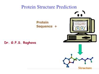
Steps to tertiary structure prediction • Comparative protein modeling • Extrapolates new structure based on related family members • Steps • Identification of modeling templates • Alignment • Model building
Identification of modeling templates • One chooses a cutoff value from FastA or BLAST search (E <10-5) • Up to ten templates can be used but the one with the highest sequence similarity to the target sequence (lowest E-value) is the reference template • Ca atoms of the templates are selected for superimposition. • This generates a structurally corrected multiple sequence alignment
Alignment • “Common core” and conserved loops of target sequence are threaded onto the template structure using only alpha carbons
Building the model • Framework construction • Average the position of each atom in target, based on the corresponding atoms in template. • Portions of the target sequence that do not match the • template are constructed from a “spare part” algorithm. • Each loop is defined by its length and C atom • coordinates of the four amino acids preceding • and following the loop.
Building the model • Completing the backbone-a library of PDB entries is consulted to add carbonyl groups and amino groups. The 3-D coordinates of the carbonyl groups and amino groups come from a separate library of pentapeptide backbone fragments. These backbone fragments are fitted onto the target C alpha carbons. • Side chains are added from a table of most probable rotamers corresponding to a particular backbone conformation. • Model refinement-minimization of energy
Protein Modeling Workshop • Continue to work through the first 11 chapters of the tutorial you began in the previous class. (http://www.usm.maine.edu/~rhodes/SPVTut/text/SPdbVTut.html) • Perform the protein modeling exercise described at: http://us.expasy.org/spdbv/text/modeling.htm