Mechanisms of TGF-β2 Expression in Trabecular Meshwork Cells
Investigating how TGF-β2 expression and release are regulated in human trabecular meshwork cells, critical for understanding glaucoma. Examining effects of lovastatin on Rho-associated signaling pathways impacting TGF-β2 synthesis and secretion.

Mechanisms of TGF-β2 Expression in Trabecular Meshwork Cells
E N D
Presentation Transcript
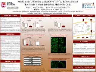
Mechanisms Governing Constitutive TGF-β2 Expression and Release in Human Trabecular Meshwork Cells Andrea L. Blitzer1, Cynthia L. Pervan-Von Zee2, Jonathan D. Lautz3, Kelly A. Langert2, and Evan B. Stubbs, Jr.2 1Stritch School of Medicine, 2Ophthalmology, 3Program of Neuroscience, Loyola University Chicago, Maywood, IL Research Service (151), Edward Hines, Jr. VA Hospital, Hines, IL Cytosol Membrane Pan-Rho RhoA RhoB GAPDH 0.01 0.1 1 0.01 0.1 1 0 10 0 10 Lovastatin (µM) A B C D B A RESULTS PUTATIVE MECHANISM INTRODUCTION Figure 1. Inhibition of geranylgeranylation attenuates endogenous TGF-β2 mRNA expression in human TM cells Figure 4. Lovastatin attenuates RhoAactivation in human TM cells Primary open angle glaucoma (POAG) is a leading cause of blindness worldwide, affecting over 2 million individuals aged 45 and over within the US. Clinically, elevated intraocular pressure (IOP) is a key risk factor for the development and progression of POAG. Harmful elevation of IOP in patients with POAG results from an increase in aqueous humor (AH) outflow resistance, largely localized to the trabecular meshwork (TM). Whereas the pathogenic mechanisms underlying elevated IOP remain poorly understood, endogenous levels of active transforming growth factor (TGF)-β2 are elevated by 60-70% in the AH of POAG patients.1-3 Experimentally, ex vivo perfusion of TGF-β2 significantly enhances outflow resistance through cultured human anterior segments.4 TGF-β2 may increase AH outflow resistance through enhanced synthesis and secretion of extracellular matrix (ECM) proteins, as well as ECM-modulating enzymes.4,5 Alternatively, we have recently shown that TGF-β2 mediated increases in AH outflow resistance may involve Rho-dependent re-organization of the actin cytoskeleton within human trabecular meshwork cells.6 The mechanisms governing TGF-β2 expression within the anterior segment of the eye remains unclear. In the healthy anterior chamber, TGF-β2 is expressed in limbal epithelial cells, the ciliary body, and the conjunctivalstroma.7In this study, we examined the mechanisms underlying endogenous synthesis and secretion of TGF-β2 by human TM cells. Anterior chamber Active TGF-β2 Transformed Primary GTP Rho hTM cell HMG-CoA HMG-CoA reductase Lovastatin Rho kinase LIM kinase Mevalonate GGTI-298 Geranylgeranyltransferase-I Mevalonate-5-P Confluent GTM3 cells were incubated in the absence (0.01% ethanol) or presence of lovastatin (10 µM), as indicated. The content of GTP-bound RhoA in cell lysates was quantified by G-LISA. Data shown are pooled from two separate experiments performed in triplicate and expressed as the mean ± SD. *p < 0.001 (unpaired Student’s t-test).10 Human TM cells were incubated for 24h in the absence (0.1% ethanol or 0.1 % DMSO) or presence of lovastatin (10 µM) or GGTI-298 (20 µM) as indicated. Relative content of TGF-β2 mRNA was quantified by qRT-PCR. Data shown are the pooled GAPDH-normalized fold changes from three separate experiments (left panel) or a single experiment (right panel) performed in triplicate and expressed as mean ± SD. *p < 0.05 (unpaired Student’s t-test). Mevalonate-5-PP Cytoskeletal reorganization Isopentenyl-PP Geranyl-PP Latent TGF-β2 METHODS Geranylgeranyl-PP (GGPP) + IPP Farnesyl-PP (FPP) GGPP synthetase Figure 5. Inhibition of geranylgeranylation disrupts endogenous filamentous actin organization in transformed human TM cells Figure 2. Inhibition of geranylgeranylation attenuates constitutive TGF-β2 secretion in human TM cells Squalene Cell Culture: An SV40-transformed human TM cell line was cultured as previously described.8 Primary human TM cells were harvested and purified from discarded human corneoscleral rims as we have previously described.6Human TM cell cultures were maintained at 37ºC in an atmosphere of 5% CO2/95% air. Treatment of Human TM Cells: Human TM cells were treated as indicated x 24h-48h in the absence (0.1% ethanol or 0.1% DMSO) or presence of lovastatin (10 µM) or GGTI-298 (10-20 µM), a geranylgeranyl transferase-I inhibitor. Lovastatin was chemically activated by alkaline hydrolysis prior to use.8 • Real-Time RT-PCR: Total RNA was extracted from human TM cells using TRIzol reagent, and 5 µg was reverse-transcribed using Super Script III First Strand Synthesis system (Life Technologies) as previously described.9Human specific TGF-β2 cDNAsequences were amplified by real-time PCR using the following primers: Sense, 5’-GCCCACTTTCTACAGACCCTACTTCAG; Antisense, 5’-GGACTTTATAGTTTTCTGAT-CACCACTGG. GAPDH (Sense, 5'-TCCCTCAAGATTGTCAGCAA; Antisense, 5-AGATC-CACAACGGATACATT) primers were used as a reference control. For each sample, the specificity of the real-time reaction product was determined by melting curve analysis. Reaction efficiencies were typically >90%. The endogenous expression of GAPDH was unaltered by drug treatments; therefore, relative fold-changes in gene expression in each sample were normalized to GAPDH. • TGF-β2 ELISA: Levels of biologically active TGF-β2 present in cell culture supernatants were assessed using a commercially-available ELISA kit. Results were read at 450 nm with a 540 nm correction and expressed as pg of active TGF-β2. • Western Immunoblotting: Proteins (20 µg per lane) from lysates of cultured cells were resolved by polyacrylamide gel electrophoresis (4-20%) and transferred to nitrocellulose membranes. Membranes were blocked and incubated overnight at 4°C in the presence of a 1:200-10,000 dilution of mouse or rabbit anti-pan-Rho, RhoA, RhoB, or GAPDH primary antibody. Washed membranes were incubated for 1h at 23oC with a 1:2,500-10,000 dilution of horseradish peroxidase-conjugated goat anti-mouse or anti-rabbit secondary antibody. Immunostainedproteins were visualized by ECL. • Rho Activation Assay:The activation of Rho was assessed by ELISA using the commercially-available G-LISA™ activation assay kit. The results were read at 490 nm. • Filamentous Actin Staining:TM cells grown to confluency on chambered coverslips were fixed (buffered 4% paraformaldehyde) and filamentous actin was stained with AlexaFluor488-conjugated phalloidin. Stained cells were visualized by confocal microscopy. Statistical Analysis:Results are expressed as mean ± SD. Parametric data were analyzed by Student’s t-test or by one-way ANOVA followed by either a Dunnett’s or Bonferroni’s multiple comparison post-hoc analysis. In all cases, p < 0.05 was considered statistically significant. Latency Associated Peptide ? Cholesterol Nucleus Transformed Primary TGF-β2 CRE/ATF E-box TATA box GCACGTCA CACGTG TATAAA +1 -74/-67 -50/-45 -30/-25 Human TM cells were treated for 24h as indicated in the absence (0.1% ethanol or 0.1% DMSO) or presence of lovastatin (10 µM) or GGTI-298 (20 µM), as indicated. Content of active TGF-β2 present in culture medium was quantified by ELISA. Data shown are from a single experiment performed in triplicate and expressed as mean ± SD. Left panel; Statistical significance is indicated (one-way ANOVA with Bonferroni’s post-hoc analysis). Dotted line represents baseline TGF-β2 content present in quiescent culture medium. Right panel; *p < 0.05 (unpaired Student’s t-test). Representative confocal images of GTM3 cells grown to confluency and treated for 24h with (A) 0.01% ethanol, (B) 10 µM lovastatin, (C) lovastatin supplemented with GGPP, or (D) 10 µM GGTI-298. Data shown are images of 2-12 independent cell preparations. Bar, 20 µm.8 Figure 3. Lovastatin elicits cytosolic accumulation of Rho GTPases CONCLUSION Figure 6. Lovastatin disrupts endogenous filamentous actin organization in primary human TM cells • TM cells represent an endogenous source of biologically active TGF-β2 within the anterior segment • Synthesis and secretion of TGF-β2 is mediated, most likely, by activation of monomeric Rho GTPase signaling • Prenyltransferase inhibitors may represent a novel therapeutic strategy for the management of glaucomatous patients with elevated IOP REFERENCES InataniM. Tanihara H, Katsuta H, Honjo M, Kido N, Honda Y. (2001) Graefe's Arch ClinExpOphthalmol239: 109-113 Min SH, Lee TI, Chung YS, Kim HK. (2006) Korean J Ophthalmol20: 162-165 PichtG, Welge-Luessen U, Grehn F, Lutjen-Drecoll E. (2001) Graefe's Arch ClinExpOphthalmol239: 199-207 Fleenor DL Shepard AR, Hellberg PE, Jacobson N, Pang IH, Clark AF. (2006) Invest OphthalmolVis Sci47: 226-234 FuchshoferR. Yu AH, Welge-Lussen U, Tamm ER. (2007) Invest Ophthalmol Vis Sci48: 715-726 Von Zee CL, Langert KA, and Stubbs EB Jr.(2012) Invest Ophthalmol Vis Sci53: 5279-5286 Pasquale LR, Dorman-Pease ME, Lutty GA, Quigley HA, Jampel HD.(1993) Invest Ophthalmol Vis Sci34:23-30 Von Zee CL Richards MP, Bu P, Perlman JI, Stubbs EB Jr. (2009) Invest Ophthalmol Vis Sci50, 2816-2823 Stubbs EB Jr. and Von Zee CL (2012) MolNeurobiol46: 28-40 Von Zee CL and Stubbs EB Jr. (2011) Invest Ophthalmol Vis Sci52: 1676-83 SUMMARY ACKNOWLEDGMENTS The authors acknowledge Charles Bouchard, M.D. for assistance with primary TM cell culture and Linda Fox for technical assistance. Supported, in part, by a Loyola University Chicago STAR award (ALB), as well as grants from the Illinois Society for the Prevention of Blindness, the Department of Veterans Affairs (B3756 (EBS)), the Midwest Eye-Banks, and the Richard A. Perritt Charitable Foundation. Confluent GTM3 cells were incubated in the absence or presence of lovastatin, as indicated (0–10 µM; 24h). Shown is a representative immunoblot from three separate experiments of pan-Rho, RhoA, and RhoB protein present in soluble (cytosol) and particulate (membrane) subcellular fractions (20 µg/lane) prepared as previously described.8GAPDH content is shown for comparison as a loading control. • Human TM cells are shown for the first time to express and secrete active TGF-β2 • Geranylgeranylated small monomeric GTPases are most likely involved in constitutive expression and release of TGF-β2 • Lovastatin prevents Rho GTPase membrane localization, while eliciting a concomitant increase in cytosolic RhoA and RhoB protein accumulation • RhoA activation was significantly attenuated by lovastatin treatment • Lovastatin disrupts filamentous actin organization in human TM cells by limiting protein geranylgeranylation Representative confocal images of primary human TM cells grown to confluency and treated for 48h with (A) 0.01% ethanol or (B) 10 µM lovastatin. Data shown are images of 2-8 independent cell preparations. Bar, 20 µm.8