Download
1 / 39
410 likes | 1.05k Vues
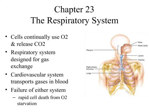
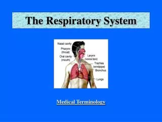
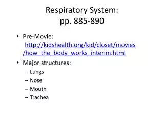
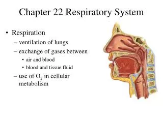
Chapter 7.1 – Structures of the Respiratory System. Pages 244 - 247. The main functions of the respiratory system: bring oxygen to cells remove carbon dioxide from cells Respiration is the general term used to describe gas exchange. M ain requirements for respiration:

E N D
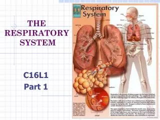
Chapter 7.1 – Structures of the Respiratory System Pages 244 - 247
The main functions of the respiratory system: • bring oxygen to cells • remove carbon dioxide from cells • Respiration is the general term used to describe gas exchange
Main requirements for respiration: • Large surface area for gas exchange • Moistenvironment allows oxygen and carbon dioxide to dissolve in water • Stages of respiration: • Breathing • Externalrespiration • Internal respiration • Cellular respiration
The Stages in Respiration • Breathing: • Inspiration – breathing in; inhaling. Moves air from the external environment to lungs inside body • Expiration – breathing out; exhaling. Moves air from lungs back to external environment
The Stages in Respiration • External respiration: exchange of oxygen and carbon dioxide between air and blood • Internal respiration: exchange of oxygen and carbon dioxide between cells and blood • Cellular respiration: energy releasing reactions taking place in the cells; sole means of providing energy for all cellular activities
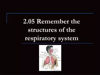
The Upper Respiratory Tract • The nasal passages: • beginning of respiratory tract • warms, moistens and cleans incoming air • Specialized cells secrete mucous and trap foreign particles
Ciliated cells sweep mucous and foreign particles back up to nose and throat where they are expelled via coughing or sneezing
Pharynx (throat): • passage way for air into the respiratory system • Epiglottis: • flap of cartilage behind tongue and larynx • closes over trachea when swallowing • prevents food from entering lungs • when epiglottis is at rest, allows air to pass into lower respiratory tract
Larynx (voice box): • made from cartilage • contains vocal cords • large gap between the vocal cords with normal breathing • larynx contracts and the vocal cords are drawn togetherwhen speaking • Air passing through narrow space causes the vocal cords to vibrate and make a sound
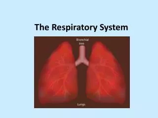
The Lower Respiratory tract • The trachea (windpipe) branches into the left and right bronchi (singular – bronchus)
Each bronchus subdivides to create branching network of bronchioles • Bronchi are supported by “C-shaped” cartilaginous rings that surround bronchus wall
Each lung divided into lobes • The right lung has 3 lobes • The left lung has 2 lobes, leaving space for heart • Each lung made up of smaller lobules extending from each bronchiole
Lungs surrounded by thin, double-layered membrane called pleural membrane • Outer layer of membrane attaches to inside of chest cavity • Inner layer of the membrane attaches to lungs • Fluid fills space between two membranes, adhering them together
Connection between the two membranes allows for lungs to expand and contract with thoracic cavity
Each bronchiole ends in a cluster of tiny sacs called alveoli (singular – alveolus)
Gas exchange occurs within alveoli during external respiration • Each alveolus is contained within a membrane called an alveolar wall • The alveolar wall is one cell thick and is surrounded by network of tiny capillaries • Alveoli are lined with lubricating film (surfactant) that prevents alveoli from collapsing
7.2 – Breathing and Respiration Pages 249 - 254
The Structures • The diaphragm and intercostal muscles (rib muscles) control movement of air in and out of lungs • Diaphragm – domed shaped muscle separating the thoracic and abdominal cavity • Intercostal muscles–found between and along ribs
The Mechanics of Breathing • Inhalation: • External rib muscles and diaphragm contract. • Rib cage expands upward and outward • The volume of the thoracic cavity increases
The Mechanics of Breathing • The density of gas in the cavity decreases – air pressure in the cavity decreases • Air moves from area high to low pressure (outside lungs to inside lungs) • Air rushes intolungs
Exhalation: External rib muscles and diaphragm relax The volume of thoracic cavity decreases The density of gas in the cavity increases – air pressure in the cavity increases Air moves from areas of high to low pressure (inside the lungs to outside the lungs) Air rushes out of the lungs
Respiratory Volume • Under normal circumstances regular breathing does not use full lung capacity • When your body needs more oxygen the volume of lungs can increase • A spirograph is used to represent the amount of air that moves into and out of lungs with each breath
The following terms are used in a spirograph: Tidal volume – volume of air inhaled and exhaled in a normal breathing movement when body is at rest Inspiratory volume –additional volume of air taken into the lungs, beyond a regular breath Expiratory reserve volume –additional volume of air that can be forced out of lungs, beyond a regular breath
Vital capacity –total lung capacity; total volume of gas that can be moved into or out of the lungs. • Vital capacity = tidal volume + inspiratory reserve + expiratory reserve volume • Residual volume –amount of air that remains in lungs and passageways after a full exhalation. If this gas left the system, the lungs and passageways would collapse. • The residual volume has little value for gas exchange because it is not exchanged with external environment
External Respiration • External respiration takes place in lungs • During external respiration, gases are exchanged between alveoli and blood in capillaries • The walls of alveoli and capillaries are each one cell thick • Gases easily diffuse through cell walls
Diffusion occurs from high low concentration The air entering alveoli have a higher oxygen concentration than blood in the capillaries, oxygen diffuses fromalveoli into capillaries
Blood in capillaries has higher concentration of carbon dioxide because it is returning from body tissues The carbon dioxide diffuses from capillaries into alveoli The carbon dioxide is exhaled into air
Internal Respiration • Once oxygen and carbon dioxide have been exchanged, the blood moves through the heartand back to body tissues • Oxygen and carbon dioxide are transported differently • 99% of oxygen is carried by hemoglobin • 1% is dissolved in blood plasma
Carbon dioxide is carried via: • 23% by hemoglobin • 7% in the plasma • 70% is dissolved and carried in the blood as bicarbonate ion (HCO3-) • The carbon dioxide reacts with water in the blood to form carbonic acid (H2CO3) • The reaction is reversed once the carbonic acid reaches the lung tissues • CO2+ H2O H2CO3 CO2+ H2O
What to do Answer the following questions on page 254 of the text: 1- 4 a,c, 6, 8
2. The typical tidal volume for humans is 500 mL. • 3. The typical expiratory reserve volume for humans is 1200 mL. • 4. The typical vital capacity for humans is 4800 mL.