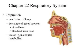
Chapter 22 Respiratory System
Chapter 22 Respiratory System. Respiration ventilation of lungs exchange of gases between air and blood blood and tissue fluid use of O 2 in cellular metabolism. Organs of Respiratory System. Nose, pharynx, larynx, trachea, bronchi, lungs. General Aspects of Respiratory System.

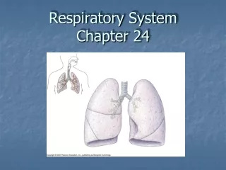
Chapter 22 Respiratory System
E N D
Presentation Transcript
Chapter 22 Respiratory System • Respiration • ventilation of lungs • exchange of gases between • air and blood • blood and tissue fluid • use of O2 in cellular metabolism
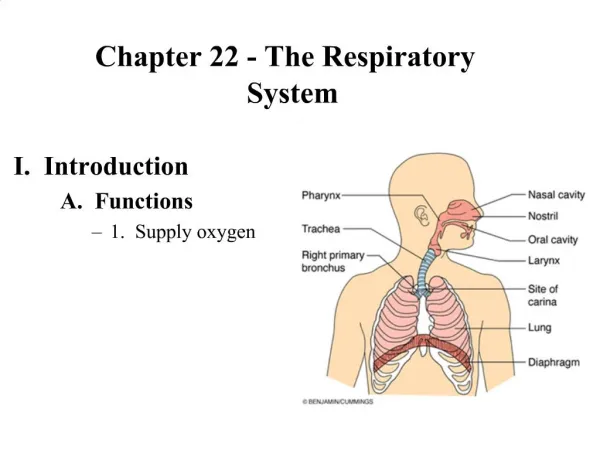
Organs of Respiratory System • Nose, pharynx, larynx, trachea, bronchi, lungs
General Aspects of Respiratory System • Airflow in lungs • bronchi bronchioles alveoli • Conducting division • passages serve only for airflow, nostrils to bronchioles • Respiratory division • alveoli and distal gas-exchange regions • Upper respiratory tract • organs in head and neck, nose through larynx • Lower respiratory tract • organs of the thorax, trachea through lungs
Nose • Functions • warms, cleanses, humidifies inhaled air • detects odors • resonating chamber that amplifies the voice • Bony and cartilaginous supports (fig. 22.2) • superior half: nasal bones medially + maxillae laterally • inferior half: lateral and alar cartilages • ala nasi: flared portion shaped by dense CT, forms lateral wall of each nostril
Pharynx • Nasopharynx (pseudostratified epithelium) • posterior to choanae, dorsal to soft palate • receives auditory tubes and contains pharyngeal tonsil • air turns 90 downward trapping large particles (>10m) • Oropharynx (stratified squamous epithelium) • space between soft palate and root of tongue, inferiorly as far as hyoid bone, contains palatine and lingual tonsils • Laryngopharynx (stratified squamous epithelium) • hyoid bone to cricoid cartilage (inferior end of larynx)
Larynx • Glottis - superior opening • Epiglottis - flap of tissue that guards glottis, directs food and drink to esophagus • Infant larynx • higher in throat, forms a continuous airway from nasal cavity that allows breathing while swallowing • by age 2, more muscular tongue, forces larynx down
Nine Cartilages of Larynx • Epiglottic cartilage • Thyroid cartilage - largest, has laryngeal prominence • Cricoid cartilage - connects larynx to trachea • Arytenoid cartilages (2) - posterior to thyroid cartilage • Corniculatecartilages (2) - attached to arytenoid cartilages like a pair of little horns • Cuneiformcartilages (2) - support soft tissue between arytenoids and the epiglottis
Walls of Larynx • Interior wall has 2 folds on each side, from thyroid to arytenoid cartilages • vestibular folds: superior pair, close glottis during swallowing • vocal cords:produce sound • Intrinsic muscles - rotate corniculate and arytenoid cartilages, which adducts (tightens: high pitch sound) or abducts (loosens: low pitch sound) vocal cords • Extrinsic muscles - connect larynx to hyoid bone, elevate larynx during swallowing
Trachea • Rigid tube 4.5 in. long and 2.5 in. in diameter, anterior to esophagus • Supported by 16 to 20 C-shaped cartilaginous rings • opening in rings faces posteriorly towards esophagus • trachealis spans opening in rings, adjusts airflow by expanding or contracting • Larynx and trachea lined with ciliated pseudostratified epithelium which functions as mucociliary escalator
Bronchial Tree • Primary bronchi (C-shaped rings) • arise from trachea, after 2-3 cm enter hilum of lungs • right bronchus slightly wider and more vertical (aspiration) • Secondary (lobar) bronchi (overlapping plates) • branches into one secondary bronchus for each lobe • Tertiary (segmental) bronchi (overlapping plates) • 10 right, 8 left • bronchopulmonary segment: portion of lung supplied by each
Bronchial Tree 2 • Bronchioles (lack cartilage) • have layer of smooth muscle • pulmonary lobule: portion ventilated by one bronchiole • divides into 50 - 80 terminal bronchioles • terminal bronchioles • have cilia , give off 2 or more respiratory bronchioles • respiratory bronchioles • divide into 2-10 alveolar ducts • Alveolar ducts - end in alveolar sacs • Alveoli - bud from respiratory bronchioles, alveolar ducts and alveolar sacs
Inspiration - Muscles Involved • Diaphragm (dome shaped) • contraction flattens diaphragm • Scalenes • fix first pair of ribs • External intercostals • elevate 2 - 12 pairs • Pectoralis minor, sternocleidomastoid and erector spinae muscles • used in deep inspiration
Smoking and Lung Cancer • Lung cancer accounts for more deaths than any other form of cancer • most important cause is smoking (15 carcinogens) • Squamous-cell carcinoma (most common) • begins with transformation of bronchial epithelium into stratified squamous • dividing cells invade bronchial wall, cause bleeding lesions • dense swirls of keratin replace functional respiratory tissue
Lung Cancer • Adenocarcinoma • originates in mucous glands of lamina propria • Small-cell (oat cell) carcinoma • least common, most dangerous • originates in primary bronchi, invades mediastinum, metastasizes quickly
Progression of Lung Cancer • 90% of lung tumors originate in primary bronchi • Tumor invades bronchial wall, compresses airway • Often first sign is coughing up blood • Metastasis is rapid and has usually occurred by time of diagnosis • common sites: pericardium, heart, bones, liver, lymph nodes and brain • Prognosis poor • 7% of patients survive 5 years after diagnosis