Download
1 / 54
540 likes | 651 Vues
Explore the origin, evidence, and implications of cell theory, including unicellular and multicellular organisms, differentiation, importance of stem cells, and size comparison of viruses, bacteria, and cells. Learn the significance of surface area to volume ratio and characteristics of prokaryotic cells.

E N D
Syllabus Statements • 2.1.1 – outline cell theory • 1.1.2 – State that a virus is a non-cellular structure consisting of DNA or RNA surrounded by a protein coat • 1.1.3 – State that all cells are formed from other cells • 1.1.4: Explain three advantages of using light microscopes • 1.1.5: Outline the advantages of using electron microscopes • 1.1.6: Define organelle • 1.1.7: Compare the relative sizes of molecules, cell membrane thickness, viruses, bacteria, organelles & cells. Use appropriate SI units. • 1.1.8: Calculate the linear magnification of drawings • 1.1.9: Explain the importance of the surface area to volume ratio as a factor limiting cell size. • 1.1.10: State that unicellular organisms carry out all of the functions of life • 1.1.11: Explain that cells in multicellular organisms differentiate to carry out specialized functions by expressing some of their genes but not others • 1.1.12: Define tissue, organ, organ system
Cell Theory • Three principles based on different studies • All organisms made of cells • Cells are the basic unit of life • Cells come from other preexisting cells • BUT… All cells aren’t created equal
Evidence of Cell theory • 1 – many organisms studied and found to consist of cells • Work of schleiden and schwann started this • Are some questionable cases – hyphae in fungi, muscle fibers, some tissues like bone • 2 – Nothing smaller than cells can “survive” when isolated • 3 – experiments show that spontaneous generation is impossible • Only evidence for cells forming from cell division
Unicellular organisms • Like amoeba, euglena etc. are made of just one cell • They do everything necessary to be called alive • Metabolism, homeostasis, response, growth, reproduction, and nutrition or energy use
Multicellular organisms • Have many cells • Allows compartmentalization of function = specialization • Multicellular Organisms therefore have emergent properties where they can do more than the sum of their parts • Take life in general being the sum of a bunch of non living chemical reactions
To have different cells do different things you need differentiation • So cells develop along different pathways or differentiate • This means different cells express different genes • Remember that every cell in an organism has all the same DNA, only some cells express different genes within that genome • Once the developmental pathway of a cell is started then it is usually fixed
So which cells can differentiate? • Stem cells – can self renew and differentiate • Human embryos are almost all stem cells • Some still found in different human tissues like skin, liver bone marrow • Those only used for limited repair
Therapeutic use of stem cells • Area of rapid development – many uses exist • Cord blood from umbilical cord contains hematopoietic stem cells – can become any blood cell type • Test the blood and remaining fluid • Used to treat some leukemias – chemo to kill the cells that over produce white blood cells then introduce cord blood to blood stream of patient • Stem cells establish themselves in the marrow and replace defective cells
How small are we talking? Viruses = 100 nm Bacteria = 1 um Organelles = up to 10 um cells = up to 100 um Chick egg = 5 cm Nerve/muscle cells in leg = 1 m Human height > 1 m
If given a scale bar • Measure the scale bar in centimeters • Measure the desired aspect of the cell in cm • Set up and solve a proportion – • Scale (um) / scale measure (cm) = actual size (um) / measured size (cm) • If given the magnification • Measure desired aspect of cell in cm • Convert your measurement to um (multiply by 10,000) • Divide by magnification to get actual size • (image size / actual size ) = magnification
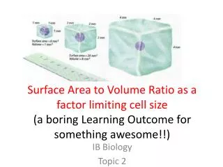
Importance of the Surface area to Volume Ratio or why are cells so small The larger the cell, the harder it is to communicate from one part to the other; more difficult to move substances through diffusion. As cell size increases, volume increases as a cubic function. Chemical reaction rate is a function of volume. Transport of necessary materials is a function of surface area increasing as a squared function.
1.2 Prokaryotic Cells • 1.2.1: Draw a generalized Prokaryotic cell • Include; cell wall, plasma membrane, mesosome, cytoplasm, ribosomes, & nucleoid (region containing naked DNA) • 1.2.2: State one function for each of the following: cell wall, plasma membrane, mesosome, cytoplasm, ribosomes, & nucleoid (region containing naked DNA • 1.2.3: State that prokaryotes show a wide range of metabolic activity including photosynthesis, fermentation, nitrogen fixation
Prokaryotes • Lack membrane-enclosed organelles • DNA is not associated with protein, chromosome is frequently circular and double stranded • Hereditary material is located in nucleoid region; no membrane separates DNA from the rest of the cell.
Functions • Cell wall = Rigid, protective layer • Plasma membrane = Regulates materials entering cell • Mesosome = folding of plasma membrane, functions in respiration • Cytoplasm = semifluid matrix of sugars, amino acids, proteins that the cell uses to carry out everyday activities • Ribosomes = protein synthesis, 70s in prokaryotes • Nucleoid (region containing naked DNA) = genetic information of the cell
When prokaryotes divide they do so by the process of binary fission
1.3 Eukaryotic Cells • 1.3.1: Draw a diagram to show the ultrastructure of a generalized animal cell as seen in an electromicrograph • Include ribosomes, rough ER, lysosome, Golgi apparatus, mitochondrion, nucleus • 1.3.2: State one function of each of the following organelles: ribosomes, rough ER, lysosome, Golgi apparatus, mitochondrion, nucleus • 1.3.3: Compare prokaryotic & eukaryotic cells • 1.3.4: Describe three differences between plant and animal cells • 1.3.5:State the composition and function of the plant cell wall
Function • Ribosomes = 80s, protein assembly • Rough ER = secretory protein & membrane synthesis, forms vessicles • Lysosomes = Digest all major classes of macromolecules, Autopathy, Apoptosis • Golgi apparatus = Center of manufacturing, warehousing, sorting, and shipping • Mitochondrion = Cellular respiration • Nucleus = contains genetic info • Chloroplast = Photosynthesis
Prokaryotes (All are Prokaryotae) Very small (1-10 um) No nucleus (nucleoid – naked DNA in central area) No membrane bound organelles – like mitochondria Have cell walls 70s ribosome Eukaryotes (all other kingdoms) Larger (10-100um) Protein asociated DNA in chromosomes in nucleus Many M.B.O. like mitochondria Plants & some fungi have cell walls 80s ribosome Prokaryoties vs. Eukaryotes
Roles of extracellular components Plant Cells • Functions to protect plant cells, maintain their shape and prevent excess water intake. • Taken together helps support plants against gravity Animal Cells • Animal cells secrete glycoproteins to form the extracellular matrix • Functions in support, adhesion, movement
1.5 Cell Division • 1.5.1: State that cell division involves interphase, mitosis & cytokinesis • 1.5.2: State that interphase is an active period in the life of a cell when many biochemical reactions occur, as well as the transcription and replication of DNA • 1.5.3: Describe the events that occur in the 4 phases of mitosis (prophase, metaphase, anaphase, telophase) • 1.5.4: Explain how mitosis produces two genetically identical nuclei • 1.5.5: Outline the difference in mitosis and cytokinesis between plants and animals • 1.5.6: State that growth, tissue repair, and sexual reproduction involve mitosis • 1.5.7: State that tumors (cancers) are the result of uncontrolled cell division and that these can occur in any organ
Why cells divide • Cell division functions in reproduction, growth and repair. • unicellular organisms: the division of one cell to form two reproduces an entire organism. – asexual reproduction Mitosis allows: • Growth • Development from fertilized egg • Replacement of dead and damaged cells
Interphase • 90% of cell cycle • A period of intense biochemical activity during which the cell grows and copies it chromosomes in preparation for cell division. • Consists of three periods: G1 phase: first growth phase S phase: synthesis of DNA; chromosomes replicate G2 phase: 2nd growth phase
Prophase • In nucleus, nucleoli disappear • Chromatin fibers condense into discrete chromosomes composed of 2 chromatids which are identical; joined at centromere. • In cytoplasm: mitotic spindle forms from cytoskeleton microtubules between 2 centrosomes. • Centrosomes move apart caused by lengthening of microtubules. • Nuclear envelope fragments
2. Metaphase • Centrosomes positioned at opposite poles of cell. • Chromosomes move to metaphase plate, long axis is at right angle to spindle axis. • Centromeres of all chromosomes aligned on the metaphase plate. • Kinetochores of sister chromatids face opposite poles.
Anaphase • Characterized by movement. Begins with paired centromeres of each chromosome move apart. • Sister chromatids split apart into separate chromosomes and move to opposite poles of the cell. • Movement is centromere first. • Kinetochore microtubules shorten at kinetochore end. • Poles of cell move father apart (elongation of cell). • At end of anaphase, 2 poles have identical collections of chromosomes.
5. Telophase • Nonkinetochore microtubules elongate the cell • Daughter nuclei begin to form at 2 poles • Nuclear envelopes form around chromosomes. • Nucleoli reappear • Chromatin fiber of each chromosome uncoils and chromosomes become less distinct. • Cytokinesis begins – 2 separate daughter cells.
Result = 2 Identical daughter cells • Interphase DNA duplicates • Chromosomes form with 2 identical chromatids joined at the centromere • Metaphase chromosomes line up on metaphase plate, kinetchore of each chromatid facing opposite poles • Anaphase separates identical chromatids (now chromosomes) into new daughter cells • By replication and organized division of genetic material the result is 2 identical daughter cells
6. Cytokinesis: division of the cytoplasm. Begins in telophase. Animal Cells • Occurs as cleavage. Cleavage furrow forms as a shallow groove in the cell surface near the old metaphase plate. • A contractile ring of actin microfilaments forms on the cytoplasmic side of the furrow. It contracts until it pinches parent cell in 2. • Remaining mitotic spindle breaks and 2 cells are completely separate.
Cytokinesis: Plant cells In plant cells, cytokinesis occurs by cell plate formation across the parent cell’s midline (old metaphase plate). • Golgi-derived vesicles move along microtubules to the cell’s center, where they fuse into a disc-like cell plate. • Additional vesicles fuse around the edge of the plate and fuse with existing parent cell’s plasma membrane. • A new cell wall forms as cellulose is deposited between the 2 membranes of the cell plate.