Role of Rho GTPases in TGF-β2-Mediated ET-1 Induction in Eye Cells
Explore the crucial role of Rho GTPases in TGF-β2 signaling leading to ET-1 expression in human trabecular meshwork cells, potentially impacting glaucoma progression.

Role of Rho GTPases in TGF-β2-Mediated ET-1 Induction in Eye Cells
E N D
Presentation Transcript
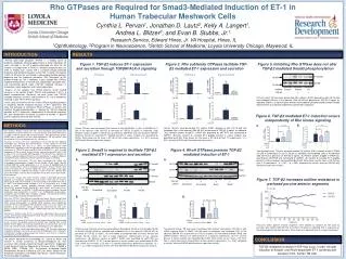
Rho GTPases are Required for Smad3-Mediated Induction of ET-1 in Human Trabecular Meshwork CellsCynthia L. Pervan1, Jonathan D. Lautz2, Kelly A. Langert1, Andrea L. Blitzer3, and Evan B. Stubbs, Jr.1Research Service, Edward Hines, Jr. VA Hospital, Hines, IL1Ophthalmology, 2Program in Neuroscience, 3Stritch School of Medicine; Loyola University Chicago, Maywood, IL INTRODUCTION RESULTS Primary open-angle glaucoma (POAG) is a leading cause of blindness worldwide, affecting approximately 2 million individuals 40 years and older in the US.A poorly-understood risk factor for the initiation and progression of POAG is elevated intraocular pressure (IOP).In healthy eyes, normal IOP is sustained through balanced production and outflow of aqueous humor (AH). In adults, the majority (>50%) of AH exits the eye through a conventional outflow pathway, beginning with the trabecular meshwork (TM).Resistance to AH outflow through the TM is mediated, in part, through Rho GTPase mediated enhancement of TM cell actin cytoskeleton contractile tone, and transforming growth factor (TGF)-β2 mediated increases in extracellular matrix deposition and F-actin organization. Analysis of AH samples from POAG patients reveal marked increases in the content of both TGF-β2 and endothelin-1 (ET-1), a potent vasoconstrictor. Previously, we have shown that TGF-β2 markedly enhances synthesis and secretion of ET-1 by a mechanism involving, in part, Rho GTPase signaling. In this study, we examine the role of Rho GTPase signaling pathways in mediating TGF-β2 mediated induction of ET-1 expression. We show that activation of monomericRhoA GTPases, in concert with selective Smad3 phosphorylation, is required for TGF-β2 mediated induction of ET-1 expression, independently of Rho kinase signaling. We also demonstrate increased resistance to outflow in porcine anterior segments perfused with TGF-β2. • Figure 3. Rho subfamily GTPases facilitate TGF-β2 mediated ET-1 expression and secretion Figure 1. TGF-β2 induces ET-1 expression and secretion through TGFβRI/ALK-5 signaling Figure 5. Inhibiting Rho GTPase does not alter TGF-β2 mediated Smad3 phosphorylation 30 kDa P~Smad2 Primary Transformed Primary Transformed 50 kDa 20 kDa RhoA Smad3 50 kDa 20 kDa RhoB Smad2 50 kDa GAPDH Primary human TM cells were pre-treated (24h) without (0.05% glycerol) or with C3 (10 µM) and incubated (30 min) in the absence (200 nMHCl) or presence of TGF-β2 (5 ng/ml) as indicated.Shown is a representative Western immunoblotof phosphorylated and total Smad3 representative of 2 separate experiments performed in duplicate. • Figure 6. TGF-β2 mediated ET-1 induction occurs independently of Rho kinase signaling METHODS Inhibitor: Vehicle Vehicle C3 B. A. _ + + TGF-β2: Human TM cells were pre-treated (1h) without (0.05% glycerol) or with C3 (10 µM) and incubated (24h) in the absence (200 nMHCl) or presence of TGF-β2 (5 ng/ml) as indicated. (Top) Relative content of ppET-1 mRNA was quantified by qRT-PCR and normalized to GAPDH. (Bottom) Content of secreted ET-1 peptide present in culture medium was quantified by ELISA. Data shown are the mean ± SD (N=3-6) from 1-2 experiments. *p < 0.01, one-way ANOVA with Dunnett’s post-hoc analysis. Human TM cells were co-treated (24h) without or with SB-431542 (1 µM) or SB-505124 (1 µM) in the absence (200 nMHCl) or presence of TGF-β2 (5 ng/ml) as indicated. (Top) Relative content of ppET-1 mRNA was quantified by qRT-PCR and normalized to GAPDH. (Bottom) Content of secreted ET-1 peptide present in culture medium was quantified by ELISA. Data shown are the mean ± SD (N=3) from single experiments. *p < 0.01, one-way ANOVA with Dunnett’s post-hoc analysis. • Cell Culture: Primary human TM cells were harvested and purified from discarded human corneoscleral rims as we have previously described (Von Zee et al., 2009). SV40-transformed TM cells derived from a male glaucomatous patient (GTM3) were a generous gift from Alcon laboratories. TM cell cultures were maintained at 37°C in an atmosphere of 5% CO2/95% air. • Treatment of Human TM Cells: Recombinant human TGF-β2 (Cell Signaling Technology) was activated and reconstituted as a stock solution as we have previously described (Von Zee et al., 2012). To pharmacologically inhibit TGFβRI/ALK-5 signaling, human TM cells were co-incubated (24h) in the absence or presence of TGF-β2 (5 ng/ml) and SB-431542 or SB-505124 (1 µM). To inhibit endogenous GTPase activity, human TM cells were pre-treated (24h) with 10 µM of either (i) C3, an irreversible ADP ribosylator of the Rho GTPase subfamily, (ii) Y-27632, a specific inhibitor of ROCK1, or (iii) Cytochalasin D, a potent inhibitor of actin polymerization, followed by TGF-β2 treatment (5 ng/ml) as indicated. • siRNA-Targeted Knockdown: TM cells were transfected with siRNA (Life Technologies) directed against Smad2 (25 nM), Smad3 (10 nM), RhoA (100 nM) or RhoB(100 nM) using Lipofectamine in a 1:1 mixture of OptiMEM and cell culture medium without serum or antibiotics/antimycotics. Primary TM cells were reverse-transfected for 8h, whereas GTM3 cells were transfected for 24h upon reaching confluence. Following transfection, culture media was replaced with serum-free medium, and cells were incubated in the absence or presence of TGF-β2 as indicated. • Western Blot: Lysates from human TM cells treated as described above were prepared in 2X Laemmli’s sample buffer and stored at -80°C until use. Proteins (20-30 µg per lane) were resolved as we have previously described (Von Zee et al., 2012), and immunostained overnight at 4°C in the presence of a 1:1,000 dilution of rabbit primary antibodies directed against: phospho-Smad2 (Ser423/425), phospho-Smad3 (Ser465/467), total Smad2, total Smad3, RhoA, RhoB (1:200 dilution), or GAPDH (1:10,000 dilution) primary antibody. Washed membranes were incubated for 1h at 23°C in a 1:10,000 dilution of peroxidase-conjugated goat-anti-rabbit IgG secondary antibody. Immunostained proteins were visualized by enhanced chemiluminescence. • Real-Time RT-PCR: Total RNA was extracted from human TM cells using TRIzol reagent and reverse-transcribed as we have previously described (Von Zee et al., 2012). Human-specific ppET-1 or GAPDH cDNA sequences were amplified by real-time PCR on a Mini-Opticon PCR detection system. For each sample, the specificity of the real-time reaction product was determined by melt curve analysis. Relative fold-changes in ppET-1 mRNA expression were normalized to GAPDH. • Endothelin-1 ELISA: Levels of ET-1 in cell culture supernatants were assessed using a commercially-available ELISA kit (R&D Systems) as we have previously described (Von Zee et al., 2012). • Porcine Anterior Segment Perfusion: Anterior segment perfusion are performed on-site at Hines VA Hospital using fresh porcine eyes obtained from a local abattoir. Porcine globes are continuously perfused at a constant flow rate of 4.5 µl/min and cultured for up to 3 additional days following pressure stabilization. • Statistical Analysis: Results are expressed as mean ± SD. Parametric data were analyzed by Student’s t-test or by one-way ANOVA followed by either a Dunnett’s or Bonferroni’s multiple comparison post-hoc analysis. In all cases, p < 0.05 was considered statistically significant. Exchange Figure 2. Smad3 is required to facilitate TGF-β2 mediated ET-1 expression and secretion Figure 4. RhoAGTPases promote TGF-β2 mediated induction of ET-1 Transformed human TM cells were pre-treated (1h) without (0.5% ethanol) or with Y-27632 (10 µM) or Cytochalasin D (Cyt D; 20 µM) as indicated, and incubated (24h) in the absence (200 nMHCl) or presence of TGF-β2 (5 ng/ml). (A) Relative content of ppET-1 mRNA was quantified by qRT-PCR and normalized to GAPDH. (B) Content of secreted ET-1 peptide present in culture medium was quantified by ELISA. Data shown are the mean ± SD (N=3-6) from 1-2 experiments. *p < 0.01; **p < 0.001 compared to vehicle,one-way ANOVA with Bonferroni’s post-hoc analysis. A. A. P~Smad3 Smad3 _ P~Smad3 • Figure 7. TGF-β2 increases outflow resistance in perfused porcine anterior segments 50 kDa + + + TGF-β2: siRNA: Scrambled RhoA RhoB _ Scrambled + + + TGF-β2: siRNA: Scrambled Smad3 Smad2 Scrambled C. B. B. 50 kDa 50 kDa Pressures within porcine anterior segments were allowed to stabilize for 24h prior to perfusion with Vehicle (400 nMHCl) or TGF-β2 (10 ng/ml) as indicated. IOP is expressed as the percentage change from baseline. Data shown are representative of a single experiment. Transformed human TM cells were transfected(24h) without (Scrambled, 100 nM) or with siRNA targeting RhoA or RhoB (100 nM each) as indicated, and incubated (24h) in the absence (200 nMHCl) or presence of TGF-β2 (5 ng/ml). (A) Immunoblot of RhoA, RhoB, and GAPDH proteins. Results are representative of 2 separate experiments. (B) ET-1 content present in culture medium was quantified by ELISA. Data shown are the mean ± SD (N=3) from a single experiment, representative of two separate experiments.*p < 0.001 compared to vehicle, one-way ANOVA with Bonferroni’s post-hoc analysis. Primary human TM cells were transfected without (Scrambled, 25 nM) or with Smad3 (10 nM) or Smad2 (25 nM) siRNA as indicatedand incubated (24h) in the absence (200 nMHCl) or presence of TGF-β2 (5 ng/ml). (A) Immunoblot of phosphorylated and total Smad3 and Smad2. Results shown are from a single experiment, representative of 2-3 separate experiments. (B) Relative content of ppET-1 mRNA was quantified by qRT-PCR and normalized to GAPDH. (C) ET-1 content present in culture medium was quantified by ELISA. Data shown are the mean ± SD from 1-2 separate experiments performed in triplicate. *p < 0.01; **, p < 0.001 compared to vehicle, one-way ANOVA with Bonferroni’s post-hoc analysis. CONCLUSION The authors would like to acknowledge Drs. Donna M. Peters and Jennifer A. Faralli (University of Wisconsin-Madison) for their assistance with anterior segment perfusion experiments. Supported, in part, by grants from the Department of Veterans Affairs (C3638R & B3756 (EBS), C7506M (CP), Pre-Doctoral Associated Health Rehabilitation Research Fellowship (CP)), the Illinois Society for the Prevention of Blindness, the Midwest Eye-Banks, and the Richard A. Perritt Charitable Foundation. TGF-β2 mediated increases in IOP may occur, in part, through induction of Smad3- and RhoA-dependent ET-1 synthesis and secretion from human TM cells.