Download
1 / 197
1.98k likes | 2.18k Vues
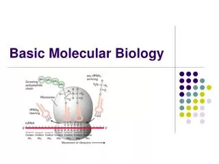
Basic Processes of Molecular Biology. Core Course # MM 702 Dr. Sonia Siddiqui Dr. Panjwani Centre For Molecular Medicine and Drug Research (PCMD). Meselson-Stahl experiment. DNA replication is Semiconservative. Bidirectional DNA replication begins at an Origin.

E N D
Basic Processes of Molecular Biology Core Course # MM 702 Dr. Sonia Siddiqui Dr. Panjwani Centre For Molecular Medicine and Drug Research (PCMD)
Meselson-Stahl experiment DNA replication is Semiconservative
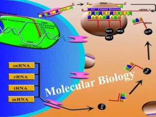
DNA Replication and Recombination Synthesis of DNA molecule: in 3 steps 1- Initiation 2- Elongation 3-Termination These processes required many different types of enzymes 1-DNA replicase system or replisome 2- Helicases 3- Topoisomerases 4- Primase 5- DNA ligases
Initiation • E.Coli DNA replication origin called OriC containing 246 bp. • For replication specific sequencing are present which is recognized by the enzymes involved in the initiation: • 1- 9 bp sequences on which DnaA protein binds • DnaA-binding sites (I sites), IHF (Integration host factor) and FIS (factor for inversion stimulation). • 2- 13 bp rich A=T sequences on which DNA unwinding element (DUE) IHF FIS
Initiation • DnaA protein is a member of the AAA+ ATPase protein family • Function: formation of oligomers and hydrolyze ATP (do things slowly) • 8 DnaA protein molecules in ATP bound form makes a helical complex emcompassing the R and I sites in oriC. • It has high affinity towards R sites than I • It binds to R sites in ATP or ADP-bound form whereas It binds to I sites in only ATP-bound form
Initiation • How DnaC docks DnaB protein? • Hexamer of each DnaC subunit bound with ATP binds with hexameric ring-shaped DnaB helicase. • This interaction of DnaB-DnaC opens DnaB ring, further interaction required DnaA • 2 out of 6 hexamer of DnaB are loaded on DUE on to each strand. • DnaC+ATP hydrolyzed, releasing DnaC and DnaB bound to the DNA.
Key step in replication: DnaB helicase docking on the DNA • DnaB unwinds the DNA from 5’→ 3’ of single stranded DNA, both strands moves in opposite direction. • This DNA with DnaB helicase has two replication forks • DNA polymerase III holoenzyme is linked via epsilon subunits • Many other single stranded DNA-binding protein (SSB) are involved that binds on each of the DNA strand at the fork • Simultaneously DNA gyrase or DNA topoisomerase II relieves the tension in the DNA molecule at the fork
The oriC DNA is methylated by Dam methylase at N6 of adenine 5’ GATC region (palindromic sequence)
Completion of DNA replication the oriC region of DNA is methylated but the newly strand is not • The hemimethylated oriC sequences are now ready to interact with the plasma membrane with the help of a protein called SeqA • OriC is released from the plasma membrane and SeqA is dissociates and DNA is fully methylated by Dam methylase
Elongation of the DNA: Leading and Lagging strand synthesis • Leading strand synthesis: • It begins with the synthesis by primase RNA primer (DnaG,10-60 nucleotide) at the fork • DnaG + DnaB helicase, primer synthesis takes place opposite in the direction of helicase movement • DnaB helicase moves along the DNA strand, the lagging strand • dNTs keep adding to the DNA strand by DNA polymerases III + DnaB complex moving on the opposite strand • Lagging strand: Okazaki fragments • On the other hand, Lagging strand synthesis starts by the formation of okazaki fragments replication direction is always from 5’- 3’ • Primase synthesize RNA primer and DNA polymerase III + DnaB adds dNTs to the lagging strand like in leading strand
It contains two subunits along with the subunits along with the ….. Subunits + AAA + ATPase • This whole complex binds to ATP and the new β sliding clamp • This creates a stretch on the dimeric clamp, opening up the ring at one subunit interface • Lagging strand slipped into the ring via breaking • Clamp loader hydrolyzes ATP, releasing the β sliding clamp Clamp loading complex of DNA polymerase III
Okazaki fragments synthesis complexity: • DNA polymerase III forms a dimer around both the strands bringing the strand close together • DnaB + DnaG complex forms at the replication fork called Replisome • DNA polymerase III has two sets of core subunits one synthesize the leading strand while the other synthesize the Okazaki fragments on the lagging strand • It is noted that at the Primosome there is β sliding clamp complex is present which is prepared by DNA polymerase III
DNA Ligases • Ligase enzyme catalyzes the formation of a phosphodiester bond between a 3’ hydroxyl at the end of one DNA strand and a 5’ phosphate at the end of another strand • Via adenylation the phosphate can be activated • Properties of DNA ligase • It is isolated from viruses and eukaryotes use ATP however, DNA ligases from bacteria are different • Many DNA ligase use NAD+ a cofactor that normally functions in hydride transfer reactions, a source of the AMP activating group • DNA ligase can also be very useful in DNA recombination experiments
Cell –Cycle control System and Activated Protein Kinases • Cyclin dependent kinases (cdks) a protein kinases which actually regulates major events of cell cycle such as DNA replication, mitosis and cytokinesis • In crease cdks levels during and at the beginning of mitosis leads to the increase phosphorylation of proteins that controls chromosome condensation, nuclear envelop breakdown and spindle assembly • However cdks activity is control by many complexes and proteins such as cyclins, cyclin activating kinases (CAK), cdk inhibitor protein (CKI), SCF, and Anaphase-promoting complex (APC), cdk25 and wee1
Cyclins-cdks complex • cdks require cyclins for their activation • Cyclins are synthesized and degraded in each cell cycle • cdks level remain normal through out the cell cycle, however changes in the levels of cyclins causes the assembly of cyclin-cdk complexes- leads to the activation and triggering of the cell-cycle events • There are four classes of cyclins G1/S-cyclins, S-cyclins, M-cyclins and G1 cyclins • Mode of activation is each complex phosphorylate the target substrate proteins and can change the activity of activation according the levels of substrate that changes during or after the cell cycle • CAK activates the cyclin-cdk complex by phosphorylating an a.a near the cdk active site—which eventually activates the target protein and induce sp cell-cycle activity
Regulation of cyclin-cdk complex • The activity of the complex can be inhibited by phosphorylation via Wee1, a protein kinase and activation can be done by a phosphatases which dephosphorylate the complex via cdc25 • The activity of the complex can be regulated by another kinases cdk inhibitor proteins (CKIs), which controls mainly S and G 1 phases . Upon binding conformational changes takes place and makes it inactive
Cyclical proteolysis and cell-cycle control system • The rate limiting step in cyclin destruction is the final ubiquitin-transfer reaction performed by 2 ubiquitin ligases, APC complex and SCF • SCF in S and G1 phase ubiquitinate the complex G1/S-cyclins and certain CKI that are involve in S phase initiation • However M phase is controlled by APC complex, it proteolyzed and ubiquitinites cyclins and other proteins involve in M phase • SCF activity is constant throughout the cell cycle, however APC levels changes with cell-cycle stages
Cell –Cycle control and Transcriptional Regulation • In more complex cell cycle, cyclins are controlled not only by there levels but by controlling at the gene transcription level and its synthesis.
Intracellular control of cell cycle events • The maintenance of each phase of cell-cycle that is G phase fusing with S phase fusing with G1 phase fusing with M phase and then G phase again, requires highly skilled and accuracy and constant adding of activating substrates to maintain the smooth overlap of the phases at different stages of cell-cycle • For eg cdc6 a regulator protein, its level increases only in G1 phase where it is required to bind with a complex with closely related proteins, minichromosomal maintenance proteins (Mcm) , resulting in the formation of a large pre-replicative complex or pre-RC complex
Intracellular control of cell cycle events • The activation of the S-cdk in late G1 initiates DNA replication, another kinases phosphorylate the Pre-Rc complex • S-Cdk helps cdc6 protein to dissociate from ORC after an origin is fired--- this leads to the disassembly of pre-RC which prevents replication from occurring again at the same origin • Secondly It prevents cdc6 and Mcm proteins from reassembling at any origin • It phosphorylates the cdc6, and triggers the ubiquitinylation by the SCF protein • S-Cdk also phoshorylates certain Mcm proteins which triggers their export from the nulceus, further proving that Mcm complex cannot bind to the replication origin • At the end all Cdk levels becomes zero, this dephosphorylate the cdc6 and Mcm proteins allow pre-Rc complex assembly to occur once again
Replication in Eukaryotes Cells Cyclin dependent kinases (CDKs) regulation control over DNA replication • The cyclins destruction by Ubiquiton-dependent proteolysis at the end of M phase • In the absence of CDKs the pre-replicative complexes (pre-RCs) can be formed on replication sites • In fast growing cells, this pre-RCs complex forms at the end M phase. Pre-RCs are called licensing • In eukaryotes the replication started by the formation of a mini chromosome maintenance (MCM) proteins • Many diff. types of MCM proteins exits like MCM2-MCM7 helicase also resembles like DnaB helicase, loads on ORC along CDC6 (cell division cycle) and CDT1 (Cell division transcript 1) • Replication requires the S phase, cyclin-cyclin dependent kinase complexes and CDC7-DBF4 • For replication both complexes must be together and the phosphorylating proteins on the pre-RCs complex
Control of replication achieved by inhibiting the synthesis of more complexes by CDK2 and other cyclins
Termination • Ter sequence trap the replication fork • Ter is for protein Tus (terminus utilization substance) binding • Ter-Tus complex works per replication cycle upon collision of either fork • Ter prevent over replication by replication fork and halts upon collision of other fork • The sequences that comes in between Ter-Tus will be replicated only , making catenane circular chromosomes
Early steps of methyl-directed mismatched repair • MutL + MutS complex at 5’ GATC binds to all mismatched base pairs • MutH + MutS binds to GATC • MutL + MutS complex creates a loop on DNA at both sides • MutH has specific endonuclease activity cleaves unmethylated GATC seq. • MutH cleaves only G at 5’ side of GATC seq.
Finishing of methyl-directed mismatched repair • When the mismatching is at 5’ • Ummethylated strand is degraded in 3’-5’ • This requires many enzymes • 1-DNAhelicase II • 2- SSB • 3- Exonuclease I OR X • 4- DNA polymerase III • 5-DNA ligase • When the mismatching is at 3’ • Exonuclease will be either VII (for degradation in to 3’-5’ or 5’-3’) OR RecJ nuclease (degrades sDNA in 5’-3’)
Mechanism of DNA Repair: Base Excision Repair • DNA glycosylases recognize the AP and abasic sites (generated by the cleavage of adenine and cytosine deamination) • Uracil DNA glycosylase removes uracil only from DNA • Enzyme recognize thymidine base from Uracil in DNA ie why DNA has thymidine and not uracil
Base-excision repair pathway • Humans have 4 types of DNA glycosylase with different specificities • Humans also has hSMUG1 which also removes U • TDG and MBD4 removes U or T present with G • Other DNA glycosylase recognize and removes formamidoprymidine and 8-hydroxyguanine (arised from purine deamination) • It also removes hypoxanthine and alkylated bases like 3-methyladenine and 7-methylguanine
Mechanism of DNA Repair:Nucleotide-Excision Repair 6th 8th 22nd 5th
Excinucleases DNA repair in E. Coli • Enzymatic complex ABC excinuclease (can create two cleavages) • Subunits: • 1- UvrA Mr 104,000 • 2- UvrB Mr 78,000 • 3- UvrC Mr 68,000
Eukaryotic excinucleasesDNA repair system : DNA damages caused by cigarette smoke can be repair by this repair mechanism • Repair mechanism like nucleotide-excision repair and base-excision repair is tied to transcription in eukaryotes • This pathway helps to repair DNA from various carcinogens like benzo[ά] pyrene-guanine, cyclobutane pyrimidine dimers and 6-4 photoproducts
Direct Repair: Damage caused by alkylating agents on nucleotide • O6-methylguanine forms in the presence of alkylating agents • This makes pairs with thymine instead of cytosine leading mismatched A-T and C-G bonds • Repairment is achieved by O6-methylguanine-DNAmethyltransferase • This enzyme transfer a methyl group of O6-methylguanine to one of its own Cys residues
Direct Repair: Damage caused by alkylating agents on nucleotide
Direct repair: Alkylated bases by AlkB • 1-methyladenine and 3-methylcytosine is repaired by ά-ketoglutarate-Fe2+ -dependent dioxygenase superfamily • In this repair A and C residues which sometimes becomes methylated in ssDNA, which affects correct base pairing • In E. coli, oxidation demethylation of these bases is mediated by AlkB protein, a member of this enzyme superfamily
Consequences of Replication fork + DNA damage • Lesion in dsDNA and ssDNA appears when the damaged DNA didn’t find complementary strand for the correct synthesis or when a replication fork encounters unrepaired DNA lesion • Error-prone translesion DNA synthesis: • The DNA repair under this pathway is less accurate • with high mutation • In bacteria this pathway is ON only when there is a • continuous damage to the cell’s DNA (oxidation or stress) • like SOS response • The production of normally present proteins UvrA and UvrB • Increases • Other proteins UmuC and UmuD activated • UmuD protein regulated by SOS response and cleaved • in to UmuD’ • UmuD’+ UmuC complex to form a specialized DNA polymerase V, helps in replication • Still difficult to make base pairing, hence can have many chances of error
Consequences of Replication fork + DNA damage • Desperate strategy from a cell to start the synthesis of UmuC and UmuD initiated by a SOS response resulting in the activation of DNA polymerase V is a deliterious. Many daughter cell dies due to the activation of this type of repair mechanism • Continuous degradation of the DNA molecule also activates RecA protein that binds ssDNA on one chromosomal location and binds with DNA polymerase V at distant sites.
Consequences of Replication fork + DNA damage • DNA polymerase η (eta) found in all eukaryotes and initiates TLS primary β, iota and λ have specialized role in base- excision repair • These enzymes also have 5’-deoxyribose PO4 lyase activity • After the removal of base by glycosylase and PO4 group by AP endonuclease, Polymerase removes the abasic site (5’ PO4) and fill in the short gap • This leads to the reduction in DNA polymerase η activity due to the short length of DNA
You tube Links- DNA Repair http://www.youtube.com/watch?v=kp0esidDr-c&feature=related http://www.youtube.com/watch?v=nPS2jBq1k48&feature=related http://www.youtube.com/watch?v=nPS2jBq1k48&feature=related http://www.youtube.com/watch?v=y16w-CGAa0Y&feature=related http://www.youtube.com/watch?v=y16w-CGAa0Y&feature=related http://www.youtube.com/watch?v=nUzyrBC0tTY http://www.youtube.com/watch?v=idbGJsDXDFo&NR=1
DNA Recombination • Homologous Genetic Recombination: Involves genetic exchange between two molecules DNA having similar sequences • Site-specific recombination: Exchange occurs only at particular sequence on a DNA • DNA transposition: Short segment of DNA in which chromosome moves from one location to another
Homologous Genetic Recombination: Base-pairing between two homologous DNA molecule • Meiosis characteristics • Two Different chromosome from two homologous DNA cross over= DNA break and ends join to their opposite partners to re-form two intact helices • Both of these helices contains half and half part of both the DNA. • The site of cross over or the exchange of the part of DNA molecules can occur anywhere in the entire DNA having homologous nt sequences in both DNA molecules
Homologous Genetic Recombination: Base-pairing between two homologous DNA molecule • This type of recombination occurs when a long region of nt sequences on both the strands are in a match • The point at which the cross over occur is called DNA synapsis • Qs arises that how both the strands recognize the site to start cross over ???
Homologous Genetic Recombination: Meiotic Recombination by dsDNA breaks • The break in PO4 diester bond attracts the other DNA helix to form base pairing thus forms a synapsis • It is thought that these strands search base pairing on another DNA strand having matching or homologous sequences • Leading to the formation of a point or joint between maternal and paternal chromosome