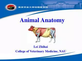
Animal Anatomy
Animal Anatomy. Lei Zhihai College of Veterinary Medicine, NAU. 14 sensory organs. 14 sensory organs.

Animal Anatomy
E N D
Presentation Transcript
Animal Anatomy Lei Zhihai College of Veterinary Medicine, NAU
14 sensory organs Sensory organs include the receptors and their accessory organs. The receptor receive the various stimuli from the external or internal enviroment of the body, and convert it into the nerve impulse. According to the position of the receptors and the origin of the stimulation, the receptors may be divided into three kinds. 1 the exteroceptors they lie in the skin, the mucous membrane of the nasal cavity, paranasal sinuses and oral cavity, and the visual and auditory organs. They receive stimuli such as touch, pain, temperature, light and sound from the external environment.
14 sensory organs 2 the enteroceptors they lie in the walls of viscera and blood vessels, and accept the physical and chemical stimuli from the internal environment. 3 the proprioceptors they lie in the muscles, tendons, joints, ligaments and the internal ear, and receive stimuli about the movement of the limbs and trunk, and the stimuli about the equilibrium.
14.1 visual organ Visual organ视觉器官 The visual organ consists of eyeball and accessory organs of the eyeball. The eyeball can receive stimulation of light and transform it into the ner- ve impulse which is conducted throu- gh the visual pathways to the visual center in the brain. The accessory or- gans include the eyelid, conjunctiva, lacrimal apparatus, extraorcular mu- scles, the conective tissue in the orbit and so on.
14.1.1 eyeball Eyeball 眼球 The eyeball lies in the orbit, embedded in the fat tissue of the orbit, and enclo- sed in a fascial sheath, posteriorly it is connected with the diencephalon by optic nerve. The eyeball is spherical in shape, however, the outline of the eyeball is not evenly rounded, but displays a larger curvature in its posterior part than in its anterior part, where the cornea bulges forward. Anterior pole Posterior pole Optic axis (external axis)
14.1.1.1 wall of eyeball Structure of the eyeball 眼球的结构 The eyeball comprises the wall of the eyeball and the contents enclosed. The wall of eyeball consists of three coats, from outside to inwards, the fibrous tunic纤维膜 sclera and cornea thevascular tunic血管膜 choroid, ciliary body and iris theretina视网膜 optic part of the retina nonvisual retina
14.1.1.1.1 fibrous tunic The fibrous tunic纤维膜, The fibrous layer of the eyeball is co- mposed of very dense collagenous ti- ssue, which is large responsible for the shape of the eye. It is composed of two parts: the opaque, whitish sclera, which enclose approximately the posterior three-quarters of the globe, and the transparent cornea, which covers the anterior part of the globe. The two components meet at the corneoscleral junction, also referred to as the corneal limbus 角膜缘.
14.1.1.1.2 vascular tunic The vascular tunic血管膜 The vascular layer of the eyeball is in- terposed between the sclera and the retina. It is composed of connective ti- ssue, which contains pigment cells, ela- stic fibers, a nervous plexus and a den- se network of blood vessels. The vascu- lar layer consists of three parts: Choroid 脉络膜 Ciliary body 睫状体 Ris 虹膜
14.1.1.1.2.1 choroid Choroid 脉络膜 It is a pigmented, highly vascular layer that envelops the posterior part of the eyeball. Dorsal to the optic papillae is a half-moon-shaped area that has an ad- ditional reflective layer, the tapetum Lucidum照膜. The tapetum lucidum is present in all domestic mammals oth-er than the pig. It aids vision during dawn and night. It has a distinctive colour in the different species and breeds (yellow in the cat, green in the dog, blue-green in the ox and horse) and accounts for the iridescent appearance of an animal’s eye.
14.1.1.1.2.2 ciliary body Ciliary body睫状体 It is the thicked middle segment of the vascular tunic, between the choroid and the iris. This is an elevated ring from which ridges, the ciliary proces- ses, radiate towards the lens in the cen- tre. Zonular fibers extend from the ciliary processes to the equator of the lens to suspend it around its periphery. The ciliary body can be subdivided into the: Ciliary ring Crown of ciliary body: contains ciliary processes Ciliary muscle: parasympathetic innervation, contract, focu-ses on near objects; sympathetic innervation, relax, distant ob.
14.1.1.1.2.3 iris Iris 虹膜 It is the most anterior part of the vas- cular layer. It is a thin ring of highly vascular tissue that rests against the anterior surface of the lens. The free margin of the iris borders the pupil, through which light enters the posterior part of the eye. The iris separates the space between cornea and lens into anterior and posterior chambers of the eye, which communicate thro-ugh the pupil. It contains two smooth muscles, the sphincter and dilator of the pupil, which regulate the size of the pupil and consequently the amount of light that reaches the retina. The sphincter muscle consists of circular smooth muscle fibe-rs near the pupillary margin of the iris.
14.1.1.1.2.3 iris The dilator of the pupil is composed of radially arranged mu-scle fibers. The pigmented cells of the iris contain melanin, which protec-ts the retina from intense light. The number and size of the pigments determines the colour of the eye. blue or grey: in pig and goat. paucity of pigmented cells. dark brown: in ox and horse. many pigmented cells. lighter, yellow: in dog, pig, small ruminants. Fewer pigmen-ted cells. red: in albinos The upper and lower pupillary margins of the iris of rumina-nts and horses show irregular outgrowths that contain coils of capillaries, the granula iridica虹膜粒, which can secrete aqueous humour.
14.1.1.1.3 retina The retina视网膜 The inner layer of the eyeball is the retina. It can be divided into the fol- lowing parts: Non-visual retina (blind part) -ciliary part of the retina and iridial part of the retina Optica part of the retina The optic part of the retina lines the internal surface of the choroid, and extends from the optic disc to the ora serrate which is the demarcation between non-visual and optic parts. In the ventrotemporal quadrant of the retina the optic nerve pierces the retina and forms the optic disc, the form of which ranges from round to oval or triangular. The optic disc is a blind point, since there are no photo-receptor cells here. loca-ted a short distance of dorsotemporal to the optic disc is an area of maximum optic resolution, the macula 黄斑.
14.1.1.2 contents of eyeball The contents of the eyeball include the aqueous humor, the lens and vitreous body. They are all transparent and ava- scular, with the cornea altogether form the refractive media. Each plays a part in refracting the light entering the eye. Aqueous humor眼房水 glaucoma青光眼 Lens 晶状体 老花眼、近视、远视、白内障 It is a transparent and elastic, biconvex body lacking blood vessels and nerves. It is surrounded by an elastic membrane known as the lens capsule and attached to the inner side of the ciliary processes by the ciliary zonule. Vitreous body玻璃体 is a colorless, transparent and jelly-like body.
14.1.2 adnexa of the eye The adnexa of the eye include the fol- Lowing accessory structures: Eyelids Lacrimal apparatus Extrinsic muscles of the eyeball Orbital fasciae Orbit with the orbital fat body Vessels and nerves
14.2 ear The ear has three subdivisions: External ear外耳 -auricle -external acoustic meatus -tympanic membrane Middle ear中耳 -tympanic cavity -auditory ossicles (hammer, anvil and stirrup) -auditory tube Internal ear内耳 -bony labyrinth -membranous labyrinth
Middle and inner ear return