Chapter 12: Cardiovascular Physiology
Chapter 12: Cardiovascular Physiology. Chapter 12: Cardiovascular Physiology. CO = HR x SV, as follows. . The heart is the pump that moves the blood. Its activity can be expressed as “cardiac output (CO)” in reference to the amount of blood moved per unit of time.


Chapter 12: Cardiovascular Physiology
E N D
Presentation Transcript
Chapter 12: Cardiovascular Physiology
Chapter 12:Cardiovascular Physiology CO = HR x SV, as follows. The heart is the pump that moves the blood. Its activity can be expressed as “cardiac output (CO)” in reference to the amount of blood moved per unit of time.
Chapter 12:Cardiovascular Physiology (cont.) A small fraction of cardiac muscle cells, called the autorhythmic cells, determine the heart rate (HR). A much larger group, making up 99% of the total cells in the heart, constitutes the contractile cells. Their activity determines the stroke volume (SV).
Chapter 12:Cardiovascular Physiology (cont.) Mean arterial pressure, which drives the blood, is the sum of the diastolic pressure plus one-third of the difference between the systolic and diastolic pressures. The autonomic system dynamically adjusts CO and MAP. Blood composition and hemostasis are described.
The hematocrit is a rapid assessment of blood composition.It is the percent of the blood volume that is composed of RBCs (red blood cells). Section :A: Overview Plasma includes water, ions, proteins, nutrients, hormones, wastes, etc. Figure 12-1
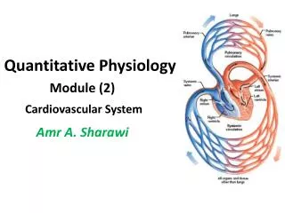
Figure 12-2 The heart is the pump that propels the blood through the systemic and pulmonary circuits. Red color indicates blood that is fully oxygenated. Blue color represents blood that is only partially oxygenated.
Figure 12-3 The distribution of blood in a comfortable, resting person is shown here. Dynamic adjustments in blood delivery allow a person to respond to widely varying circumstances, including emergencies.
Figure 12-4 Though pressure is higher in the lower “tube,” the flow rates in the pair of tubes is identical because they both have the same pressure difference (90 mm Hg) between points P1 and P2.
Section :B: The major external and internal parts of the heart are shown in this diagram. The black arrows indicate the route taken by the blood as it is pumped along. Figure 12-6
Figure 12-8 The general route of the blood through the body is shown, including passage through the heart (colored box).
Figure 12-9 Cardiac muscle structure
Conducting system of the heart Figure 12-10
Sequence of cardiac excitation Figure 12-11 The Bundle of His and other parts of the conducting system deliver the excitation to the apex of the heart so that ventricular contraction occurs in an upward sweep. The sinoatrial node is the heart’s pacemaker because it initiates each wave of excitation with atrial contraction.
The rapid opening of voltage-gated sodium channels is responsible for the rapid depolarization phase. The action potential of a myocardial pumping cell. Figure 12-13
The action potential of a myocardial pumping cell. The prolonged “plateau” of depolarization is due tothe slow but prolonged opening of voltage-gated calcium channels PLUS closure of potassium channels. Figure 12-12
The action potential of a myocardial pumping cell. Opening of potassium channels results in the repolarization phase. Figure 12-12
The action potential of a myocardial pumping cell. The rapid opening of voltage-gated sodium channels is responsible for the rapid depolarization phase. The prolonged “plateau” of depolarization is due tothe slow but prolonged opening of voltage-gated calcium channels PLUS closure of potassium channels. Opening of potassium channels results in the repolarization phase. Figure 12-12
Figure 12-13 The action potential of anautorhythmic cardiac cell. Sodium ions “leaking” in through the F-type [funny] channels PLUS calcium ions moving in through the T [calcium]channels cause a threshold graded depolarization. The rapid opening of voltage-gated calcium channels is responsible for the rapid depolarization phase. Reopening of potassium channels PLUS closing of calcium channels are responsible for the repolarization phase.
Figure 12-14 The relationship between the electrocardiogram (ECG), recorded as the difference between currents at the left and right wrists, and an action potential typical of ventricular myocardial cells.
Einthoven’s triangle Figure 12-15a
Normal Partial block Complete block Figure 12-16
Excitation contraction coupling in cardiac muscle Calcium ions regulate the contraction of cardiac muscle: the entry of extracellular calcium ions causes the release of calcium from the sarcoplasmic reticulum, the source of about 95% of the calcium in the cytosol. Figure 12-17
Refractory period of cardiac muscle The prolonged refractory period of cardiac muscle prevents tetanus, and allows time for ventricles to fill with blood prior to pumping. Figure 12-18
Mechanical events of the cardiac cycle Systole: ventricles contracting and ejection Figure 12-19a
Diastole: ventricles relaxed and filling Figure 12-19b
Figure 12-20 Pressure and volume changes in the left heart during a contraction cycle.
Pulmonary circulation pressures Pressure changes in the right heart during a contraction cycle. Figure 12-21
Heart Sounds Figure 12-22
The cardiac output: control of heart rate Figure 12-23
Figure 12-24 • To speed up the heart rate: • deliver the sympathetic hormone, epinephrine, and/or • release more sympathetic neurotransmitter (norepinephrine), and/or • reduce release of parasympathetic neurotransmitter (acetylcholine).
Frank-Starling mechanism To increase the heart’s stroke volume: fill it more fully with blood. The increased stretch of the ventricle will align its actin and myosin in a more optimal pattern of overlap. Figure 12-25
Stroke volume and sympathetic nerves To further increase the stroke volume: fill it more fully with blood AND deliver sympathetic signals (norepinephrine and epinephrine); it will also relax more rapidly, allowing more time to refill. Figure 12-26
Effects of sympathetic stimulation on ventricular contraction and relaxation Figure 12-27 Sympathetic signals (norepinephrine and epinephrine) cause a stronger and more rapid contraction and a more rapid relaxation.
Factors determining the cardiac output To increase SV, increase:end-diastolic volume,norepinephrine delivery from sympathetic neurons, and epinephrine delivery from the adrenal medulla. To increase HR, increase: norepinephrine delivery from sympathetic neurons, and epinephrine delivery from adrenal medulla (reduce parasympathetic). Figure 12-28 It is not possible, under normal circumstances, to increase one but not the other of these determinants of cardiac output.
Section C: The vascular system
Pressures in the systemic vessels Figure 12-29
The blood moved in a single heart contraction stretches out the arteries, so that their recoil continues to push on the blood, keeping it moving during diastole. Figure 12-30
Figure 12-32 To estimate systolic and diastolic pressures, pressure is released from an inflatable cuff on the upper arm while listening as blood flow returns to the lower arm.
Arterioles Dynamic adjustments in the blood distribution to the organs is accomplished by relaxation and contraction of circular smooth muscle in the arterioles. Figure 12-33
Local control of organ blood flow Active hyperemia and flow autoregulation differ in their cause but both result in the production of the same local signals that provoke vasodilation. Figure 12-34
Figure 12-35 Sympathetic stimulation of alpha-adrenergicreceptors cause vasoconstrictionto decrease blood flow to that location. Sympathetic stimulation of beta-adrenergic receptors lead to vasodilation to cause an increase in blood flow to that location.