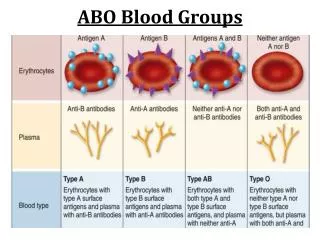
Other Blood Groups
Other Blood Groups. The Kell Blood Group System. Background information. The Kell blood group system was discovered in 1946. Number of Kell antigens: > 20 These antigens are the third most potent, after those of the ABO and Rh blood groups, at triggering an immune reaction.

Other Blood Groups
E N D
Presentation Transcript
Background information • The Kell blood group system was discovered in 1946. • Number of Kell antigens: > 20 • These antigens are the third most potent, after those of the ABO and Rh blood groups, at triggering an immune reaction.
Molecular information • The KEL gene is found on chromosome 7 • The KEL gene is highly polymorphic, with different alleles at this locus encoding the 25 antigens that define the Kell blood group. • The Kell protein is a polypeptide chain of 732 amino acids in length that becomes glycosylated at five different sites. It makes a single pass through the RBC membrane.
Kell Blood Group System • XK gene produces Kx substance, which is a precursor of of Kell Ags • Kel genes convert Kx substance into the Kell Ags on RBCs • K (Kell) & k (cellano) are produced by allelic genes, this results into 3 phenotypes: • K+k- (genotype KK) • K+k+ (genotype Kk) • K-k+ (genotype kk) • Other allelic genes include: Kpa/Kpb, Jsa/Jsb
XK Gene (Chromosome X) KEL Gene RBC Kell system glycoprotein: Kell Ag’s reside here. Kx
Kx Substance • Kx substance is present on RBCs & WBCs • Kell genes convert Kx substance into the Kell Ags on RBCs • Kell genes do not convert Kx on WBCs
McLeod Phenotype • Absence of Kx proteins in RBCs membrane lead to McLeod Phenotype • This absence cause: • abnormal RBCs shape (acanthocytes) & reduced in-vivo survival.
Chronic Granulomatous Disease • Absence of Kx proteins in WBCs cause CGD • Leukocytes are able to phagocytose but not to kill bacteria • Patients with CGD have recurrent bacterial infections • Patients who lack Kx on RBCs & WBCs have both Mcleod and CGD
Kell Null (K0) Phenotype 1.K0 is a silent Kell allele 2. When homozygous K0K0 inherited no Kell system antigens are expressed. 3. Kx antigen expression is enhanced 4. Very rare Kx
Kell Antibodies • K- individuals produce anti-K when exposed to K+ cells • Frequency of K is low (9%), easy to find compatible blood for the patient with anti-k. • On the other hand frequency of k antigen is 99.9% • Difficult to find blood
Duffy Blood Group System • The Duffy blood group was discovered in 1950. • The Duffy glycoprotein is encoded by the FY gene, found on chromosome 1 , of which there are two main alleles, FYA and FYB. They are codominant. • The Duffy gene codes for a glycoprotein also found in other tissues: brain, kidney, spleen, heart and lung. • The Duffy glycoprotein is a transmembrane protein • Five alleles at Duffy locus, the most important: Fya, Fyb & Fy (Silent Allele) • Fya is more immunogenic than Fyb
Different genes • Fy(a-b-) blacks do not produce anti-Fya oranti-Fyb following transfusion with Fy(a+) or Fy(b+) blood • Fy(a-b-) Caucasians become sensitizedfollowing transfusion with Fy(a+) or Fy(b+) blood • This suggest that Fy(a-b-) phenotype arises from different genes in the two populations
Duffy Antigens • Fya, Fyb antigens are Destroyed by enzymes • Abs DO NOT agglutinate enzyme treated cells • Moderately immunogenic.
Duffy Antibodies • IgG antibodies and can activate complement • Anti- Fya is more frequently encountered • Anti- Fyb is more frequently found in patients produced multiple alloantibodies
Duffy and Malaria • Black people with the Duffy phenotype of Fy(a–b–) appear to have resistance to Plasmodium vivax & Plasmodium knowlesi causative agents of Malaria. • Duffy antigens appear to be a receptor for the P. vivax organism and when the antigen is not present on the red blood cell membrane P. vivax is unable to access the red blood cell • Some area’s of West Africa are 100% Fy(a–b–). • Plasmodium falciparumbinds to RBCs at integral glycophorin A & B
Kidd Blood Group System • The Kidd blood group was discovered in 1950. • The Kidd gene is located on chromosome 18 • Three alleles: Jka, Jkb, Jk • Codominant Inheritance • Jk is a silent allele (amorph) • The Kidd protein is an integral protein of the RBC membrane.
Kidd Antigens & Antibodies • Ags are well developed at birth • Have tendency to drop to low or undetectable levels following formation. • Abs are of IgG type & can activate complement (Anti-Jka, Anti-Jkb ) • Produced following transfusion or pregnancy • Can cause HDNB • They are also a very common cause of delayed HTRs
Ii Blood Group • Found nearly on all RBCS • Their products aretransferase enzymes that attach repeating units of Gal and GlcNAc to the ABO Precursor Substance. • Big I gene codes for branching of the Precursor Substance.
Ii Antigens • Little i antigen is LINEAR • Found on cord cells, predominantly • Big I antigen is BRANCHED • Gradually convert from i to I during the first 18 months of life. Not all i converted to I, some i still present on adult cells, normally. • Rare adult individuals termed iadult do not express i Ag on their red cells • The I and i antigen sites are considered uncompleted ABH active chains. • When ABH are removed from RBCs more I Ags are expressed • I structure located beneath the ABH Ags
I Antibodies: Anti-I • Anti-I is naturally occurring often due to a Mycoplasma pneumoniae infection • Anti-I reacts with all adult cells (including patient’s own, all reagent cells, all donor cells) • Anti-I does not react with cord cells • Auto-anti-I is a common “cold agglutinin”
Antii Antibodies • Antii is rarely found in healthy individuals • Reacts preferably with cord cells • anti-i can be found secondary to Infectious Mononucleosis. • Transient: Only present with active disease
MNSs Blood Group System • The antigens M and N are produced by co-dominant alleles • closely linked to the S and s genes, which are also co-dominant. • Chromosome 4 contains these linked genes • Genes produce two distinct glycophorins or sialyglycoproteins (SGP) on the RBC membrane.
MN Genetics • MN Locus genes produce Glycophorin A (GPA) • M-GPA’s 1st five aa’s = Serine-Ser-Thr-Thr-Glycine • N-GPA’s 1st five aa’s = Leucine-Ser-Thr-Thr-Glutamic acid • Amino acids (aa) 2, 3 & 4 are the same for both • Glycophorin A (GPA) is a glycoprotein also known as MN-sialoglycoprotein
Ss Genetics • Ss genes code for the production of Glycophorin B(GPB) • S glycophorin B has Methionine aa at position 29 • s glycophorin B has Threonine aa at position 29 • Glycophorin B (GPB) is a glycoprotein also known as Ss-sialoglyprotein
Ss Genotypes & Phenotypes • U antigen is a high incident antigen NOT seen in individuals who lack both S and s antigens. • Individuals who lack this antigen (<1%) have a high likelihood of forming anti-U as well as anti-S and anti-s.
Anti-M Antibodies • Variability of reactivity (Dosage) • Strong reactions with RBCs homozygous for MM • Weak reactions with RBCs heterozygous MN
Anti-N antibodies • Naturally occurring cold agglutinin • Can form in patients with renal Failure • During dialysis with formaldehyde sterilized equipment • Formaldehyde may alter the N Ag structure making it appear foreign
P Blood Group System • Genetics: These genes code for enzymes that sequentially add sugars to precursor substance. • This system is related to the ABO, Le and Ii systems. • Genes: P1, Pk, P and lower case p (silent allele) • All antigens are expressed on glycolipids on red cells
Phenotypes, Detectable Antigens & Frequencies • Pk is the precursor of P. • Rare individuals do not convert Pk into P. • Those will have Pk on RBCs.
Anti-P1 Antibodies Naturally occcurring Abs found in the serum of P2 Individuals
Allo Anti-PAntibodies Naturally occcurring Abs found in the serum of Pk and pIndividuals
Auto anti-PAntibodies • It is an IgG biphasic Ab associated with Paroxysmal Cold Hemoglobinuria (PCH) • Binds complement at cold temperatures and activates that complement in warm temperatures lysing the red blood cells.
Anti TjaAntibodies • Combination of anti-P, anti-P1 & anti-Pk • Found in serum of individuals who have no P, P1 & PkAgs on red cells