Download
1 / 52
520 likes | 546 Vues
Understand the composition, structure, and function of bone tissue, bone cells, histology, and importance of calcium in the body. Explore the complexities of long bone anatomy and the differences between compact and spongy bone.

E N D
Bones and Skeletal Tissues H. Biology II Adapted 2014-2015
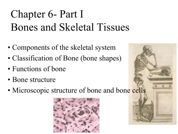
INTRODUCTION • Bone is made up of several different tissues working together: bone, cartilage, dense connective tissue, epithelium, various blood forming tissues, adipose tissue, and nervous tissue. • Each individual bone is an organ; the bones, along with their cartilages, make up the skeletal system.
The Skeletal System: Bone Tissue • Dynamic and ever-changing throughout life • Skeleton composed of many different tissues • cartilage, bone tissue, epithelium, nerve, blood forming tissue, adipose, and dense connective tissue
Functions of Bone • Supporting & protecting soft tissues • Attachment site for muscles making movement possible • Storage of the minerals, calcium & phosphate -- mineral homeostasis • Blood cell production occurs in red bone marrow (hemopoiesis) • Energy storage in yellow bone marrow
Importance of Ionic Calcium in the Body • Calcium is necessary for: • Transmission of nerve impulses • Muscle contraction • Blood coagulation • Secretion by glands and nerve cells • Cell division
Anatomy of a Long Bone • diaphysis = shaft • epiphysis = one end of a long bone • metaphyses are the areas between the epiphysis and diaphysis and include the epiphyseal plate in growing bones. • Articular cartilage over joint surfaces acts as friction reducer & shock absorber • Medullary cavity = marrow cavity
Anatomy of a Long Bone • Endosteum = lining of marrow cavity • Periosteum = tough membrane covering bone but not the cartilage • fibrous layer = dense irregular CT • osteogenic layer = bone cells & blood vessels that nourish or help with repairs
Histology of Bone • A type of connective tissue as seen by widely spaced cells separated by matrix • Matrix of 25% water, 25% collagen fibers & 50% crystalized mineral salts • 4 types of cells in bone tissue
HISTOLOGY OF BONE TISSUE • Bone (osseous) tissue consists of widely separated cells surrounded by large amounts of matrix. • The matrix of bone contains inorganic salts, primarily hydroxyapatite and some calcium carbonate, and collagen fibers. • These and a few other salts are deposited in a framework of collagen fibers, a process called calcification or mineralization. • The process of calcification occurs only in the presence of collagen fibers. • Mineral salts confer hardness on bone while collagen fibers give bone its great tensile strength.
Bone Cells • Osteogenic cells undergo cell division and develop into osteoblasts. • Osteoblasts are bone-building cells. • Osteocytes are mature bone cells and the principal cells of bone tissue. • Osteoclasts are derived from monocytes and serve to break down bone tissue.
Cells of Bone • Osteoprogenitor cells ---- undifferentiated cells • can divide to replace themselves & can become osteoblasts • found in inner layer of periosteum and endosteum • Osteoblasts--form matrix & collagen fibers but can’t divide • Osteocytes ---mature cells that no longer secrete matrix • Osteoclasts---- huge cells from fused monocytes (WBC) • function in bone resorption at surfaces such as endosteum
Matrix of Bone • Inorganic mineral salts provide bone’s hardness • hydroxyapatite (calcium phosphate) & calcium carbonate • Organic collagen fibers provide bone’s flexibility • their tensile strength resists being stretched or torn • remove minerals with acid & rubbery structure results • Bone is not completely solid since it has small spaces for vessels and red bone marrow • spongy bone has many such spaces • compact bone has very few such spaces
Compact Bone • Compact bone is arranged in units called osteons or Haversian systems . • Osteons contain blood vessels, lymphatic vessels, nerves, and osteocytes along with the calcified matrix. • Osteons are aligned in the same direction along lines of stress. These lines can slowly change as the stresses on the bone changes.
Compact or Dense Bone • Looks like solid hard layer of bone • Makes up the shaft of long bones and the external layer of all bones • Resists stresses produced by weight and movement
Histology of Compact Bone • Osteon is concentric rings (lamellae) of calcified matrix surrounding a vertically oriented blood vessel • Osteocytes are found in spaces called lacunae • Osteocytes communicate through canaliculi filled with extracellular fluid that connect one cell to the next cell • Interstitial lamellae represent older osteons that have been partially removed during tissue remodeling
Spongy Bone • Spongy (cancellous) bone does not contain osteons. It consists of trabeculae surrounding many red marrow filled spaces. • It forms most of the structure of short, flat, and irregular bones, and the epiphyses of long bones. • Spongy bone tissue is light and supports and protects the red bone marrow.
The Trabeculae of Spongy Bone • Latticework of thin plates of bone called trabeculae oriented along lines of stress • Spaces in between these struts are filled with red marrow where blood cells develop • Found in ends of long bones and inside flat bones such as the hipbones, sternum, sides of skull, and ribs. No true Osteons.
Blood and Nerve Supply of Bone • Periosteal arteries • supply periosteum • Nutrient arteries • enter through nutrient foramen • supplies compact bone of diaphysis & red marrow • Metaphyseal & epiphyseal aa. • supply red marrow & bone tissue of epiphyses
BONE FORMATION • All embryonic connective tissue begins as mesenchyme. • Bone formation is termed osteogenesis or ossification and begins when mesenchymal cells provide the template for subsequent ossification. • Two types of ossification occur. • Intramembranous ossification is the formation of bone directly from or within fibrous connective tissue membranes. • Endochondrial ossification is the formation of bone from hyaline cartilage models.
Stages of Intramembranous Ossification • Forms some flat bones of the skull, mandible, and clavicle. • An ossification center appears in the fibrous connective tissue membrane • Bone matrix is secreted within the fibrous membrane • The matrix surrounds the cell and then calcifies as the osteoblast becomes an osteocyte. • The calcifying matrix centers join to form bridges of trabeculae that constitute spongy bone with red marrow between. • On the periphery the mesenchyme condenses and develops into woven bone and the periosteum.
Stages of Intramembranous Ossification Figure 6.7.1
Stages of Intramembranous Ossification Figure 6.7.2
Stages of Intramembranous Ossification Figure 6.7.3
Stages of Intramembranous Ossification Figure 6.7.4
Endochondral Ossification • Endochondrial ossification involves replacement of cartilage by bone and forms most of the bones of the body • The first step in endochondrial ossification is the development of the cartilage model. • Begins in the second month of development • Uses hyaline cartilage “bones” as models for bone construction • Requires breakdown of hyaline cartilage prior to ossification
Endochondral Bone Formation • Development of Cartilage model • Mesenchymal cells form a cartilage model of the bone during development • Growth of Cartilage model • in length by chondrocyte cell division and matrix formation ( interstitial growth) • in width by formation of new matrix on the periphery by new chondroblasts from the perichondrium (appositional growth)
Stages of Endochondral Ossification • Formation of bone collar • Cavitation of the hyaline: cartilage cells in mid-region burst and change pH triggering calcification and chondrocyte death • Invasion of internal cavities by the periosteal bud, and spongy bone formation • Formation of the medullary cavity; appearance of secondary ossification centers in the epiphyses • Ossification of the epiphyses, with hyaline cartilage remaining only in the epiphyseal plates
Stages of Endochondral Ossification cont. • Development of Primary Ossification Center • perichondrium lays down periosteal bone collar • nutrient artery penetrates center of cartilage model • periosteal bud brings osteoblasts and osteoclasts to center of cartilage model • osteoblasts deposit bone matrix over calcified cartilage forming spongy bone trabeculae • osteoclasts form medullary cavity
Stages of Endochondral Ossification Cont. • Development of Secondary Ossification Center • blood vessels enter the epiphyses around time of birth • spongy bone is formed but no medullary cavity • Formation of Articular Cartilage • cartilage on ends of bone remains as articular cartilage.
Stages of Endochondral Ossification Secondary ossification center Articular cartilage Epiphyseal blood vessel Spongy bone Deteriorating cartilage matrix Hyaline cartilage Epiphyseal plate cartilage Spongy bone formation Primary ossification center Medullary cavity Bone collar Blood vessel of periosteal bud Formation of bone collar around hyaline cartilage model. 1 Cavitation of the hyaline cartilage within the cartilage model. 2 Invasion of internal cavities by the periosteal bud and spongy bone formation. 3 Formation of the medullary cavity as ossification continues; appearance of secondary ossification centers in the epiphyses in preparation for stage 5. 4 Ossification of the epiphyses; when completed, hyaline cartilage remains only in the epiphyseal plates and articular cartilages 5 Figure 6.8
Postnatal Bone Growth- Timeline • Growth in length of long bones • Cartilage on the side of the epiphyseal plate closest to the epiphysis is relatively inactive • Cartilage abutting the shaft of the bone organizes into a pattern that allows fast, efficient growth • Cells of the epiphyseal plate proximal to the resting cartilage form three functionally different zones: growth, transformation, and osteogenic
Long Bone Growth and Remodeling Figure 6.10
Appositional Growth of Bone Central canal of osteon Periosteal ridge Penetrating canal Periosteum Artery Osteoblasts beneath the periosteum secrete bone matrix, forming ridges that follow the course of periosteal blood vessels. As the bony ridges enlarge and meet, the groove containing the blood vessel becomes a tunnel. 1 The periosteum lining the tunnel is transformed into an endosteum and the osteoblasts just deep to the tunnel endosteum secrete bone matrix, narrowing the canal. 2 As the osteoblasts beneath the endosteum form new lamellae, a new osteon is created. Meanwhile new circumferential lamellae are elaborated beneath the periosteum and the process is repeated, continuing to enlarge bone diameter. 3 4 Figure 6.11
Factors Affecting Bone Growth • Nutrition • adequate levels of minerals and vitamins • calcium and phosphorus for bone growth • vitamin C for collagen formation • vitamins K and B12 for protein synthesis • Sufficient levels of specific hormones • during childhood need insulinlike growth factor • promotes cell division at epiphyseal plate • need hGH (growth), thyroid (T3 &T4) and insulin • sex steroids at puberty • At puberty the sex hormones, estrogen and testosterone, stimulate sudden growth and modifications of the skeleton to create the male and female forms.
Factors that affect Bone Growth • EXERCISE: Within limits, bone has the ability to alter its strength in response to mechanical stress by increasing deposition of mineral salts and production of collagen fibers. • Removal of mechanical stress leads to weakening of bone through demineralization (loss of bone minerals) and collagen reduction. • reduced activity while in a cast • astronauts in weightless environment • bedridden person • Weight-bearing activities, such as walking or moderate weightlifting, help build and retain bone mass.
Bone Remodeling • Remodeling units – adjacent osteoblasts and osteoclasts deposit and resorb bone at periosteal and endosteal surfaces
Bone Remodeling • Remodeling is the ongoing replacement of old bone tissue by new bone tissue. • Old bone is constantly destroyed by osteoclasts, whereas new bone is constructed by osteoblasts. • In orthodontics teeth are moved by braces. This places stress on bone in the sockets causing osteoclasts and osteoblasts to remodel the sockets so that the teeth can be properly aligned • Several hormones and calcitriol control bone growth and bone remodeling
Bone Remodeling • Ongoing since osteoclasts carve out small tunnels and osteoblasts rebuild osteons. • osteoclasts form leak-proof seal around cell edges • secrete enzymes and acids beneath themselves • release calcium and phosphorus into interstitial fluid • osteoblasts take over bone rebuilding • Continual redistribution of bone matrix along lines of mechanical stress • distal femur is fully remodeled every 4 months
Bone Deposition • Occurs where bone is injured or added strength is needed • Requires a diet rich in protein, vitamins C, D, and A, calcium, phosphorus, magnesium, and manganese • Alkaline phosphatase is essential for mineralization of bone • Sites of new matrix deposition are revealed by the: • Osteoid seam – unmineralized band of bone matrix • Calcification front – abrupt transition zone between the osteoid seam and the older mineralized bone
Bone Resorption • Accomplished by osteoclasts • Resorption bays – grooves formed by osteoclasts as they break down bone matrix • Resorption involves osteoclast secretion of: • Lysosomal enzymes that digest organic matrix • Acids that convert calcium salts into soluble forms • Dissolved matrix is transcytosed across the osteoclast’s cell where it is secreted into the interstitial fluid and then into the blood
Hormonal Regulation of Bone Growth During Youth • During infancy and childhood, epiphyseal plate activity is stimulated by growth hormone • During puberty, testosterone and estrogens: • Initially promote adolescent growth spurts • Cause masculinization and feminization of specific parts of the skeleton • Later induce epiphyseal plate closure, ending longitudinal bone growth
Control of Remodeling • Two control loops regulate bone remodeling • Hormonal mechanism maintains calcium homeostasis in the blood • Mechanical and gravitational forces acting on the skeleton
Hormonal Mechanism • Rising blood Ca2+ levels trigger the thyroid to release calcitonin • Calcitonin stimulates calcium salt deposit in bone • Falling blood Ca2+ levels signal the parathyroid glands to release PTH • PTH signals osteoclasts to degrade bone matrix and release Ca2+ into the blood
Hormonal Mechanism Figure 6.12
Hormonal Mechanism • Parathyroid hormone (PTH) is secreted if Ca+2 levels falls • PTH gene is turned on & more PTH is secreted from gland • osteoclast activity increased, kidney retains Ca+2 and produces calcitriol • Calcitonin hormone is secreted from parafollicular cells in thyroid if Ca+2 blood levels get too high • inhibits osteoclast activity • increases bone formation by osteoblasts
Calcium Homeostasis & Bone Tissue • Skeleton is a reservoir of Calcium & Phosphate • Calcium ions involved with many body systems • nerve & muscle cell function • blood clotting • enzyme function in many biochemical reactions • Small changes in blood levels of Ca+2 can be deadly (plasma level maintained 9-11mg/100mL) • cardiac arrest if too high • respiratory arrest if too low
Development of Bone Tissue • Both types of bone formation begin with mesenchymal cells • Mesenchymal cells transform into chondroblasts which form cartilage OR • Mesenchymal cells become osteoblasts which form bone Mesenchymal Cells
Developmental Aspects of Bones • Mesoderm gives rise to embryonic mesenchymal cells, which produce membranes and cartilages that form the embryonic skeleton • The embryonic skeleton ossifies in a predictable timetable that allows fetal age to be easily determined from sonograms • At birth, most long bones are well ossified (except for their epiphyses)
Developmental Aspects of Bone Tissue 5th Week =limb bud appears as mesoderm covered with ectoderm 6th Week = constriction produces hand or foot plate and skeleton now totally cartilaginous 7th Week = endochondral ossification begins 8th Week = upper & lower limbs appropriately named