Download
1 / 34
340 likes | 464 Vues
This project presents a novel micro-Pillar Array Detector (mPAD) designed by Richard Besen, Albert Leung, Feng Yu, and Yan Zhao under Professor Horacio Espinosa, aimed at measuring cellular adhesion forces. The mPAD consists of discrete individual force sensors modeled as cantilever beams, allowing for precise and direct calculations of adhesion forces. By optimizing material selection and pillar geometry, the device can be tailored for varying cell types, offering high sensitivity in detecting nano-Newton forces. This study addresses the challenges in adhesion force measurements and proposes a practical solution.

E N D
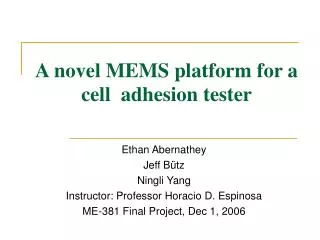
Geometrically Optimized mPAD Device for Cell Adhesion Professor Horacio Espinosa – ME 381 Final Project Richard Besen Albert Leung Feng Yu Yan Zhao Fall 2006
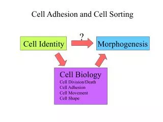
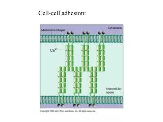
Introduction Cellular Adhesion Force • For a cell to move, it must adhere to a substrate and exert traction • Traction forces are concentrated at focal points between the cell and substrate Cellular Functions Biological Mechanism ME 381
Cellular Adhesion Video ME 381
Literature Review Adhesion Force Measurement Continuous Substrate Method Wrinkle Method • Sensitive to nano-Newton forces • Force calculations difficult because of complexity of wrinkle pattern • Model does not show adhesion force focal points ME 381
Literature Review Adhesion Force Measurement Continuous Substrate Method Gel imbedded with fluorescent markers • Highly sensitive to adhesion forces • Markers aid in optical detection of surface deformation • Difficult to manufacture uniform fluorescent marker pattern ME 381
Proposed Design Adhesion Force Measurement mPADs (micro Pillar Array Detectors) • Discrete individual force sensors • Direct calculations from cantilever deflection theory • Highly detailed force vector field • Precise and simple manufacturing ME 381
Proposed Design Adhesion Force Measurement Customization mPAD design depends on the type of cell being used Variable Parameters: • Material Selection • Aspect ratio • Pillar density • Cell to pillar contact area ME 381
Proposed Design Adhesion Force Measurement mPAD Sensing • Pillar is modeled as a cantilever beam with uniform diameter • Pillar geometry, quantity of pillars per area, material choice can be modified to match known ranges of a cell’s adhesion force • Force vector field shows magnitude and direction of discrete forces exerted by the cell on the array ME 381
Geometric and Mechanical Analysis • Force and Displacement • Area Percentage ME 381
Geometric and Mechanical Analysis • Bending Stress • Bending Moment H ME 381
Optimization • Material: • Flexible to cell adhesion forces • Optically measurable displacements • Geometry and Spatial Arrangement: • Minimize cell flow down sides of posts • Detailed vector field representation • Manufacturable ME 381
Optimization Criterion • Maximization of post density • Minimization of spring constant ME 381
Optimization Theory • Cost function: • Optimization Problem: • Lagrangean: C1, C2- Weighting Coefficients subject to ME 381
Constraints • System Dynamics: • Material: • Properties: • Yield Stress: ME 381
Height (H) 4 μm -150 μm Diameter (D) 100 nm – 5 μm Distance between posts (L) >2Δmax Constraints continued • Spatial & Geometric Parameters: • Optical Resolution: R=50nm ME 381
Optimization trends Density as a function of diameter holding height constant at 4m ME 381
Optimization trends continued Density as a function of the distance between adjacent posts holding diameter constant at 1.2141 m ME 381
Optimization trends continued Spring constant as a function of diameter holding height constant at 4m ME 381
Optimization trends continued Spring constant as a function of post height holding diameter constant at 1.2141m ME 381
Optimization trends continued Spring constant as a function of distance between adjacent posts where K=2Fmax/L and Fmax=10nN ME 381
Results ME 381
Materials • PDMS - polydimethylsiloxane • Desirable chemical, physical, and economic properties ME 381
Chemical Properties • Cell friendly • Chemically inert • Thermally stable • Non-toxic • Can be made hydrophilic for adhesion purposes ME 381
Physical Properties • Extremely flexible • (.87MPa < E < 3.6MPa) • Scalability • Conforms to nano-scale structures • Necessary for micro-molding • Transparent within visible spectrum • Cheap! Around $50 per pound to process • Adjustable stiffness and aspect ratio based on mixing ratio and curing time ME 381
Mask Oxide Si substrate UV light Mask and pattern 1 μm photoresist using UV lithography Mask 1 – quartz plate with 800Å chromium layer Photoresist Transfer pattern to mask oxide with HF isotropic etching Microfabrication Deposit mask oxide with LPCVD (SiO2) ME 381
Passivation oxide Deposit .3 μm passivation oxide with PECVD After vertical oxide etch, deep Si etch alternating with passivation Microfabrication (cont’d) • Bosch Process First deep anisotropic silicon etch (DRIE) with Cl2/BCl3 ME 381
mono-Si base substrate Cured PDMS structure soft bonded to mono-silicon substrate (E ~ 100 GPa), removed from mold Microfabrication (cont’d) • Micromolding Liquid PDMS prepolymer Liquid PDMS poured into silanized micromold ME 381
Defects • Scalloping from imperfect etch selectivity in DRIE (~100 nm) • Variable diameter (conic shape) ME 381
Preparation and Fluorescent Labeling • Oxidize structure in air-plasma to make hydrophilic • Create flat PDMS stamps for top of each pillar • Microcontact print fluorescent label • Coat pillars and stamps in adhesive ME 381
mPAD Calibration • Spring Constant (K) • AFM Curves • Young’s Modulus (E) • Compression • Height/Diameter • SEM analysis ME 381
Optical Sensing • Phase-Contrast Microscopy • Epifluroescence Microscopy ME 381
Optical Sensing (cont’d) • Pillar Deflection Detection • Force Analysis Package ME 381
Future Studies • 3D Analysis – Software improvements ME 381
Thank You! • Questions? ME 381