Download
1 / 1
10 likes | 225 Vues
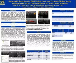
Sonographic Detection of a Bifid Median Nerve and Persistent Median Artery Among Patients with a Clinical Diagnosis of Carpal Tunnel Syndrome Francis Luk , Carolyn R. O’Connor, Humaira Hussain , Vincent J Zarro and Angel Checa Drexel University College of Medicine, Philadelphia, PA.

E N D
Sonographic Detection of a Bifid Median Nerve and Persistent Median Artery • Among Patients with a Clinical Diagnosis of Carpal Tunnel Syndrome • Francis Luk, Carolyn R. O’Connor, HumairaHussain, Vincent J Zarro and Angel Checa • Drexel University College of Medicine, Philadelphia, PA Background pma Rheumatologists often manage patients with carpal tunnel syndrome (CTS) by corticosteroid injections with or without ultrasound guidance. Although new imaging techniques that permit the identification of the bifid median nerve (BMN) and persistent median artery (PMA) are being used more frequently, the prevalence of these variants continues to be unclear. Both PMA and BMN, if present, are potential sources of complications due to laceration during interventional procedures such as carpal tunnel release or steroid injection. BMN UA A C pma Purpose B D E To determine the prevalence and sonographic characteristics of BMN and PMA in patients with carpal tunnel syndrome Figure 3. Panels A and B show power Doppler scan in short- and long-axis views, respectively. (C) Cross-sectional measurement of lateral medial branch of a BMN. (D) Power Doppler scan of a patient with a persistent median artery. (E) Transverse view of the same patient shown in (D), illustrating sonographic-guided injection in the opposite side of the PMA (arrow). BMN, bifid median nerve; pma, permanent median artery; ua, ulnar artery Methods B A total of 129 wrists of 77 consecutive patients (9 males, 68 females) with a clinical diagnosis of CTS assessed by ultrasound in a rheumatology office were studied from January 2010 to June 2011. These patients had symptoms of pain, paresthesia, and/or weakness. Longitudinal and transverse scans of the volar aspect of the wrist were performed with a GE Logiq e machine equipped with a broadband linear transducer from 8 to 13 MHz. The cross-sectional area of the median nerve was calculated with the ellipse at the scaphoid-pisiform level. In those cases with BMN, both branches were measured separately. The functional status of the median artery was evaluated by the presence or absence of a Doppler signal. A Results Of 77 patients (129 wrists), 13 (14 wrists) had BMN or PMA. The mean age of those with BMN or PMA was 52.5 (31-81) years; the prevalence of BMN and PMA was 8.5% and 4.6%, respectively. BMN was more common in the left hand, and a greater lateral branch was the most characteristic sonographic pattern, observed in 70%. The average cross-sectional area for BMN was 11.4 (range 6-18) mm2. The PMA was associated with a BMN in three wrists, and it appeared as an isolated variant in 3 other wrists. The average diameter of a PMA was 1.4 (range 0.8-3.1) mm. In three of them, the Doppler signal showed a functional artery. A Doppler signal was absent in two PMAs; in one artery, the Doppler signal was not explored. In those patients with PMA but no Doppler signal, a nonfunctional, cord-like remnant may be present. Conclusions ma mn Although studies of large populations are needed, the increased observation of BMN and PMA by ultrasound in patients with CTS should be considered more than a medical curiosity. This study demonstrates that BMN were observed in 8.5% of wrists referred for evaluation of hand pain. The PMA was seen in 4.6% of wrists. We suggest that ultrasound evaluation of the carpal tunnel should be routine procedure in the rheumatology office prior to corticosteroid injection and surgical release. ua l m cb A B C Figure 1. (A) Diagram showing the transverse orientation of the transducer. (B) Transverse sonographic view of the volar aspect of the wrist. This view shows a bifid median nerve and the nerve structures, carpal bones, and ulnar artery. (C) Magnified image of the two branches of a bifid nerve, medial and lateral, accompanied by the median artery. cb, carpal bones; l, lateral; m, medial; ma, median artery; mn, bifid median nerve; ua, ulnar artery m mn mn References l m mn B l A 1. Bayrak IK, Bayrak AQ, Kale M, Turker H, Diren B. Bifid median nerve in patients with carpal tunnel syndrome. J Ultrasound Med 2008;27:1129-1136. 2. Beris AE, Lykissas MG, Kontogeorgakos V, Vekris M, Korompilias A. Anatomic variations of the median nerve in carpal tunnel release. Clin Anat 2008;21:514-518 3. Checa A, Hussain H. Sonographic assessment of a bifid median nerve and median artery in carpal tunnel syndrome. J Rheumatol 2011;38:1694-1696. 4. Pierre-Jerome C, Smitson RD Jr, Shah RK, Moncayo V, Abdelnoor M, Terk MR. MRI of the median nerve and median artery in the carpal tunnel: prevalence of their anatomical variations and clinical significance. Surg Radiol Anat 2010;32:315-322. Short axis C D Long axis Long axis m Figure 2. (A) Bifid median nerve without the median artery. (B) Diagrammatic representation of a longitudinal sonographic scan and examination of the medial and lateral branches emerging from a common trunk. (C) Medial branch of the bifid nerve in longitudinal view. (D) Lateral branch of the bifid nerve in longitudinal view. l, lateral; m, medial; mn, bifid median nerve b l b B