Membrane rest potential . Generation and radiation action potential .
380 likes | 656 Vues
Membrane rest potential . Generation and radiation action potential. This discussion will focus on selected examples of transport catalysts for which structure/function relationships are relatively well understood. Transporters are of two general classes: carriers and channels .

Membrane rest potential . Generation and radiation action potential .
E N D
Presentation Transcript
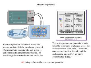
Membrane rest potential. Generation and radiation action potential.
This discussion will focus on selected examples of transport catalysts for which structure/function relationships are relatively well understood. Transporters are of two general classes: carriers and channels. These are exemplified by two ionophores (ion carriers produced by microorganisms): valinomycin (a carrier) gramicidin(a channel).
Puckering of the ring, stabilized by H-bonds, allows valinomycin to closely surround a single unhydrated K+ ion. Six oxygen atoms of the ionophore interact with the bound K+, replacing O atoms of waters of hydration. Valinomycin is highly selective for K+ relative to Na+. The smaller Na+ ion cannot simultaneously interact with all 6 oxygen atoms within valinomycin. Thus it is energetically less favorable for Na+ to shed its waters of hydration to form a complex with valinomycin.
Whereas the interior of the valinomycin-K+ complex is polar, the surface of the complex is hydrophobic. This allows valinomycin to enter the lipid core of the bilayer, to solubilize K+ within this hydrophobic milieu. Crystal structure (at Virtual Museum of Minerals & Molecules).
Valinomycin is a passive carrier for K+. It can bind or release K+ when it encounters the membrane surface. Valinomycin can catalyze net K+ transport because it can translocate either in the complexed or uncomplexed state. The direction of net flux depends on the electrochemical K+ gradient.
Proteins that act as carriers are too large to move across the membrane. They are transmembrane proteins, with fixed topology. An example is the GLUT1 glucose carrier, in plasma membranes of various cells, including erythrocytes. GLUT1 is a large integral protein, predicted via hydropathy plots to include 12 transmembrane a-helices.
Carrier proteinscyclebetweenconformations in which a solute binding site is accessible on one side of the membrane or the other. There may be an intermediate conformation in which a bound substrate is inaccessible to either aqueous phase. With carrier proteins, there is never an open channel all the way through the membrane.
The transport rate mediated by carriers is faster than in the absence of a catalyst, but slower than with channels. A carrier transports one or few solute molecules per conformational cycle, whereas a single channel opening event may allow flux of many thousands of ions. Carriers exhibit Michaelis-Mentenkinetics.
Classes of carrier proteins Uniport (facilitated diffusion) carriers mediate transport of a single solute. An example is the GLUT1glucose carrier. The ionophore valinomycin is also a uniport carrier.
Symport (cotransport) carriers bind two dissimilar solutes (substrates) & transport them together across a membrane. Transport of the two solutes is obligatorily coupled. A gradient of one substrate, usually an ion, may drive uphill (against the gradient) transport of a co-substrate. It is sometimes referred to as secondary active transport. E.g: glucose-Na+ symport, in plasma membranes of some epithelial cells bacterial lactose permease, a H+symport carrier.
Lactose permease catalyzes uptake of the disaccharidelactose into E. coli bacteria, along with H+, driven by a proton electrochemical gradient. It is the first carrier protein for which an atomic resolution structure has been determined. Lactose permease has been crystallized with thiodigalactoside (TDG), an analog of lactose.
The substrate binding site is at the apex of an aqueous cavity between two domains, each consisting of six trans-membrane a-helices. In the conformation observed in this crystal structure, the substrate analog is accessible only to what would be the cytosolic side of the intact membrane. Residues essential for H+ binding are are also near the middle of the membrane.
As in simple models of carrier transport based on functional assays, the tilt of transmembrane a-helices is assumed to change, shifting access of lactose & H+ binding sites to the other side of the membrane during the transport cycle.
Antiport (exchange diffusion) carriers exchange one solute for another across a membrane. • Usually antiporters exhibit "ping pong" kinetics. A substrate binds & is transported. Then another substrate binds & is transported in the other direction. • Only exchange is catalyzed, not net transport. The carrier protein cannot undergo the conformational transition in the absence of bound substrate.
Example of an antiport carrier: Adenine nucleotide translocase (ADP/ATP exchanger) catalyzes 1:1 exchange of ADP for ATP across the inner mitochondrial membrane.
Active transport enzymes couple net solute movement across a membrane to ATP hydrolysis. An active transport pump may be a uniporter or antiporter. ATP-dependent ion pumps are grouped into classes, based on transport mechanism, genetic & structural homology.
P-class ion pumpsare a gene family exhibiting sequence homology. They include: • Na+,K+-ATPase, in plasma membranes of most animal cells is an antiport pump. It catalyzes ATP-dependent transport of Na+ out of a cell in exchange for K+ entering. • (H+, K+)-ATPase, involved in acid secretion in the stomach is an antiport pump. It catalyzes transport of H+ out of the gastric parietal cell (toward the stomach lumen) in exchange for K+ entering the cell.
P-class pumps (cont): • Ca++-ATPases, in endoplasmic reticulum (ER) and plasma membranes, catalyze ATP-dependent transport of Ca++ away from the cytosol, into the ER lumen or out of the cell. Some evidence indicates that these pumps may be antiporters, transporting protons in the opposite direction. Ca++-ATPase pumps function to keep cytosolic Ca++ low, allowing Ca++ to serve as a signal.
The reaction mechanism for a P-class ion pump involves transient covalent modification of the enzyme. At one stage of the reaction cycle, phosphate is transferred from ATP to the carboxyl of a Glu or Asp side-chain, forming a “high energy” anhydride linkage (~P). At a later stage in the reaction cycle, the Pi is released by hydrolysis.
The ER Ca++ pump is called SERCA: Sarco(Endo)plasmic Reticulum Ca++-ATPase. In this diagram of SERCA reaction cycle, conformational changes altering accessibility of Ca++-binding sites to the cytosol or ER lumen are depicted as positional changes. Keep in mind that SERCA is a large protein that maintains its transmembrane orientation.
Reaction cycle: 1.2Ca++ bind tightly from the cytosolic side, stabilizing the conformation that allows ATP to react with an active site aspartate residue. 2.Phosphorylation of the active site aspartate induces a conformational change that • shifts accessibility of the 2 Ca++ binding sites from one side of the membrane to the other, & • lowers the affinity of the binding sites for Ca++.
3.Ca++ dissociates into the ER lumen. 4. Ca++ dissociation promotes • hydrolysis of Pi from the enzyme Asp • conformational change (recovery) that causes Ca++ binding sites to be accessible again from the cytosol.
This X-ray structure of muscle SERCA (Ca++-ATPase) shows 2 Ca++ ions (colored magenta) bound between transmembrane a-helices in the membrane domain. Active site Asp351, which is transiently phosphorylated during catalysis, is located in a cytosolic domain, far from the Ca++ binding sites.
SERCA structure has been determined in the presence & absence of Ca++, with & without inhibitors. • Substantial differences inconformation have been interpreted as corresponding to different stages of the reaction cycle. • Large conformational changes in the cytosolic domain of SERCA are accompanied by deformation & changes in position & tilt of transmembrane a-helices. • The data indicate that whenCa++ dissociates: • water molecules enter Ca++ binding sites • charge compensation is provided by protonation of Ca++-binding residues.
This simplified cartoon represents the proposed variation in accessibility & affinity of Ca++-binding sites during the reaction cycle. Only 2 transmembrane a-helices are represented, and the cytosolic domain of SERCA is omitted.
More complex diagrams & animations have been created by several laboratories, based on available structural evidence. E.g.: animation(lab of D. H. MacLennan) diagram (by C. Toyoshima, in a website of the Society of General Physiologists - select Poster). websiteof the Toyoshima Lab (select Resources for movies). websiteof the Stokes Lab (select Download movie).
Ion Channels Channels cycle between open & closed conformations. When open,a channel provides acontinuous pathway through the bilayer, allowing flux of many ions. Gramicidin is an example of a channel.
Gramicidin is an unusual peptide, with alternating D & L amino acids. In lipid bilayer membranes, gramicidin dimerizes & folds as a right-handed b-helix. The dimer just spans the bilayer. Primary structure of gramicidin (A): HCO-L-Val-Gly-L-Ala-D-Leu-L-Ala-D-Val-L-Val-D-Val-L-Trp-D-Leu-L-Trp-D-Leu-L-Trp-D-Leu-L-Trp- NHCH2CH2OH Note: The amino acids are all hydrophobic; both peptide ends are modified (blocked).
The outer surface of the gramicidin dimer, which interacts with the core of the lipid bilayer, is hydrophobic. Ions pass through the more polar lumen of the helix. Ion flow through individual gramicidin channels can be observed if a small number of gramicidin molecules is present in a lipid bilayer separating 2 compartments containing salt solutions.
With voltage clamped at some value, current (ion flow through the membrane) fluctuates. Each fluctuation, attributed to opening or closing of one channel, is the same magnitude. The current increment corresponds to current flow through a single channel (drawing - not actual data).
Gating (opening & closing) of a gramicidin channel is thought to involve reversible dimerization. An open channel forms when two gramicidin molecules join end to end to span the membrane. This model is consistent with the finding that at high [gramicidin] overall transport rate depends on [gramicidin]2.
Channels that are proteins Cellular channels usually consist of large protein complexes with multiple transmembrane a-helices. Their gating mechanisms must differ from that of gramicidin. Control of channel gating is a form of allosteric regulation. Conformational changes associated with channel opening may be regulated by: • Voltage • Binding of a ligand (a regulatory molecule) • Membrane stretch (e.g., via link to cytoskeleton)
Patch Clamping The technique of patch clamping is used to study ion channel activity. A narrow bore micropipet may be pushed up against a cell or vesicle, and then pulled back, capturing a fragment of membrane across the pipet tip.
Patch Clamping A voltage is imposed between an electrode inside the patch pipet and a reference electrode in contact with surrounding solution. Current is carried by ions flowing through the membrane.
If a membrane patch contains a single channel with 2 conformational states, the current will fluctuate between 2 levels as the channel opens and closes. Theincrement in currentbetween open & closed states reflectsthe rate of ion flux through one channel. View a videoof an oscilloscope image during a patch clamp recording.
Patch clamp recording at -60 mV. Consecutive traces are shown. Note that at a negative voltage, increased current is a downward deflection.
Current Amplitude Histogram Occupancy of different current levels during the time period of a recording is plotted against current in picoAmperes (10-12 Amp). Peaks represent open & closed states (note scale). Baseline current, when the channel is closed, is due to leakage of the patch seal and membrane permeability.