Download
1 / 55
560 likes | 840 Vues
Vascular Neck Trauma. Case 1. Presentation to Lithgow. 19M, riding motorcycle in the bush- helmet, no leathers Felt sudden sharp severe pain in R anterolateral neck Brought by friends to Lithgow Hospital Entry wound over anterolateral R SCM near angle of mandible, neck swelling

E N D
Presentation to Lithgow • 19M, riding motorcycle in the bush- helmet, no leathers • Felt sudden sharp severe pain in R anterolateral neck • Brought by friends to Lithgow Hospital • Entry wound over anterolateral R SCM near angle of mandible, neck swelling • CT neck Lightgow-metallic FB 9mmR neck, parapharyngealhaematoma with tracheal deviation • Therefore arranged for urgent transfer to Trauma Centre- Westmead Hospital
Westmead Hospital- Primary Survey • Airway: • Speaking in sentences, hoarse voice. No stridor/resp distress. • Trachea and uvula deviated to left. • No subcut emphysema or crepitus • No drooling/odynophagia/dysphagia • Zone 3 R sided puncture wound over SCM • B: SaO2 100% RA, equal air entry, normal RR, no respiratory distress
Primary Survey (cont.) • C: HR 97, BP 180/70; non-expanding non-pulsatile R neck swelling in SCM, no bruit heard • D: GCS 15/15, vocal hoarseness and deviated uvula, moving all limbs spontaneously, no focal neurological deficits, no other cranial nerve abnormalities
Secondary Survey • Head, neck, face: findings as above; no other injuries seen; no cervical spine tenderness • Chest: No chest tenderness, equal AE, vesicular breath sounds • Abdomen: soft, non-tender • Pelvis: stable and non-tender • Upper & lower limbs: NAD
Evaluation • Zone 3 penetrating neck trauma (above angle of mandible) • Potential airway compromise due to extrinsic haematoma • Moderate-high risk for vascular neck injury due to location of entry wound and haematoma • No sign of acute life threatening vascular compromise (exsanguination/haemorrhage/stroke)
Management • Urgent assessment of airway • No stridor or respiratory distress • Nasendoscopy performed by ENT: • Oropharyngeal haematoma with mild swelling • Normal vocal cords & movement • Normal cranial nerves • No need for immediate intubation, if any deterioration for anaesthetic r/v and gaseous intubation • Deemed stable for transfer to CT angiography with medical escort
Management (cont) • IV dexamethasone to minimise airway oedema • O2 therapy via Hudson mask • 2x large bore cannulae; 1L of Hartmann’s administered intravenously; analgesia • ADT and cephazolin administered
Imaging report • 2x metallic foreign bodies- one at level of C2, one embedded in SCM • 6mm ECA pseudoaneurysm 2.5cm above angle of mandible
Further management • Admission to ICU for airway, circulatory and neuro observations • Vascular consultation • Aspirin • Semi-electively 3-4 days post injury R Cerebral & carotid angiogram for management of pseudoaneurysm with coiling performed. • No immediate complications; d/c home on oral antibiotics
Presentation to WMH- Major Trauma Call • 58M awoken by partner stabbing his R neck with kitchen knife • Walk in to ED • Major trauma call on arrival
Primary Survey • Airway: • Speaking in sentences • No stridor; no tracheal deviation • 2cm laceration upper zone 2 over R SCM with small non-pulsatile non-expanding haematoma • No active bleeding • No crepitation/emphysema • No dysphagia/odynophagia/drooling • Breathing: • SaO2 95%, equal air entry, vesicular breath sounds, no respiratory distress
Primary Survey (cont) • C: HR 80, BP 140/85, small haematoma at area of stab wound • D:GCS 15/15, moving all limbs spontaneously, no focal neurological deficits, no cranial nerve abnormalities
Secondary Survey • Head, neck, face: findings as above; no other injuries seen; no cervical spine tenderness • Chest: No chest tenderness, equal AE, vesicular breath sounds • Abdomen: soft, non-tender • Pelvis: stable and non-tender • Upper & lower limbs: NAD
Evaluation • Zone 2 penetrating neck trauma (between cricoid cartilage and angle of mandible) • Stable from airway/breathing/circulatory perspective • Potential injury to anterior neck vasculature • Deemed safe for transfer for CT angiogram of head and neck
Management • 6L O2 via Hudson Mask • 2x large bore cannulae, IV Hartmann’s solution • IV cephazolin, ADT • NBM • CT angiogram of head & neck performed
Imaging report • 26mm x 20mm x 15mm subcutaneous haematoma anterolateral to R SCM superficial to inferior aspect of parotid gland • Small locule of gas in R SCM • Vessels intact
Further Management • HDU admission for airway, circulation observations • For exploration of neck wound with ASU and vascular team early the next day
Operative Findings • Expanding R anterior neck haematoma- evacuated • Stab wound tract explored- penetration through platysma to lacerated sternocleidomastoid belly • Dissection to R IJV- intact • R ICA, vagus nerve, identified-intact
Further Progress • Returned to HDU postoperatively for airway & circulatory monitoring • No immediate postoperative complications • Discharged the next day on oral antibiotics
Vascular Neck Injuries 25% of head/neck trauma 5-10% all arterial injury Carotid injury- 10-30% mortality; 15-60% permanent neurologic deficit
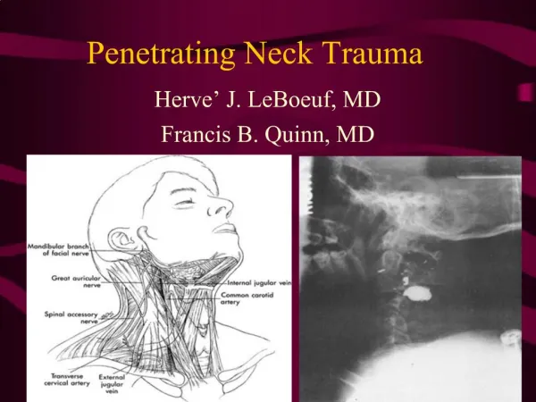
Relevant Anatomy ICA, ECA Jugular vv Lat pharynx Cr VII, IX, X, XI, XII CCA ICA, ECA Jugular vv Larynx Hypopharynx Cr X, XI, XII Subclaa & vv Jugular vv CCA Trachea Oesophagus, thyroid
Vascular traumatic injuries • Complete or partial transection • Intimal flap/dissection • Aneurysm • Pseudoaneurysm • Fistula • Extrinsic compression • Thromboembolism as a result of intimal injury
Sequelae • Haemorrhage • Airway compression, exsanguination, concealed haematoma • Distal ischaemia • Either due to vessel injury or thromboembolism • Strokes- ACA/MCA (carotid injury), PCA/posterior (vertebral injury) • Damage to nearby structures
Penetrating neck injury (>90%) • Injuries through platysma indicate propensity for injury to deep structures • Gunshot wounds and projectiles • Low velocity- unpredictabletrajectory • High velocity • Cavitation and blunt type injury from concussive forces • Stab/knife • Straight and more obvious path • Less tissue damage
Blunt Neck Trauma (<10%) • Seatbelt injury • Hanging/ligature/strangulation • Punching/kicking • Hyperextension/hyperrotation/contusion • Mechanism is translocational & shear forces • Spectrum from intimal injury (more common) to transection (less common)
Associated with dislocation/fracture • Mandibular, temporal bone fractures can be a/w carotid/jugular injury • Vertebral aa injury in general rare- usually a/w C-spine pathology • #C-spine (inc Lateral mass #) • Ligamentous injury • Rotation/hyperextension • Near-hanging • Extreme chiropractic manoevres
Iatrogenic injury • CVC insertion • Cerebral Angiography • C-spine surgery, transsphenoidal, skull base surgery • Radiotherapy (stenosis) • Nerve blocks (vertebral aa injury)
Comorbid injuries • Airway – pharynx, larynx, trachea • Pneumothorax, haemothorax (Zone 1) • Nerve injuries • Cranial VII, IX, X, XI, XII • Brachial plexus • Cervical sympathetic chain (Horner’s) • C-spine, mandibular, temporal fractures • Oesophagus • Parotid, salivary glands, lymph nodes • Thyroid (Zone 1)
Airway • High comorbidity with airway injury & compromise • Assess for: • Airway patency- stridor, resp distress, hoarseness • Expanding haematoma • Emphysema/crepitus/drooling/dysphagia • ENT r/v if possible (+/- nasendoscopy) • May require trache(/cricothyroidotomy/intubation), exploration or stenting • If unstable will require emergent OT +/- trache
Breathing • General principles apply • Give Supplemental O2 • Optimise tissue O2 delivery • Assess chest expansion & for subcut emphysema • Need CXR • May have comorbid chest injury in high risk mech (eg MVA) • Zone 1- risk of assochaemo/pneumothorax • Index of suspicion for aspiration
Circulation • General principles of resuscitation apply • Large bore IV access • Fluid resuscitation, Xmatch, possible transfusion • Direct compression of severe external bleeding- finger/foley catheter in wound • If unstable – immediate OT
Circulation (cont) • Assess for “Hard” signs of vascular injury • Pulsatile bleeding or haematoma • Expanding haematoma • Shock + ongoing bleeding • Absent pulses • Neurovascular symptoms- stroke/TIA symptoms • Thrills, bruits
Circulation (cont) • “Soft” signs – warrant further investigation • Severe bleeding from neck/pharynx • Diminished pulses- superficial temp artery • Small haematoma • Fractures of skull base, temporal bone, fracture d/location C-spine • Injury in anatomical area • Ipsilateral Horner’s • Cranial IX-XII dysfunction • Widened mediastinum
Disability • If suspicion of C-spine injury- hard collar • Focal neurology in stroke territoryshould alert to possible vasc injury • Cranial nerve VII --> XII (except VIII) • Horner’s syndrome (compression of cervical chain) • Brachial plexus injury
Other Injuries on Secondary Survey • Aerodigestive – oesophagus & pharynx • Drooling • Odynophagia, dysphagia
Summary • Airway injury/compromise common and may r/q emergent management • If unstable from airway/circulatory point of view needs immediate operative management including exploration • Expanding haematoma may cause airway compromise • Stroke symptoms, bruits, thrills are a hard sign of vascular injury • If stable can go on to have further imaging
Bloods • Hb, haematocrit (blood gas or formal) • BSL- must optimise O2 & glucose delivery • ABG in airway/breathing compromise
Plain radiography • CXR & neck XR • Foreign bodies • Injury to lung apices- haemo/pneumothorax • Mediastinal widening • Surgical emphysema, aerodigestive injuries • (C-spine fractures)
Scanning • Duplex USS useful for Zone 2 injuries- unhelpful for Z1 or 3 • CT brain & CTA neck • CT angiogram may show aneurysm, dissection, fistulae etc (esp with blunt trauma) or occult injury • Localisation of FB • CT brain valuable predictor of outome- infarct on CTB has high mortality, poor neurologic prognosis
Management Endovascular, operative, supportive
Supportive/preop care • Nurse in HDU environment • Supplemental O2 • Fluid resuscitation • Correct hypoglycaemia • Anticoagulation for intimal injuries- high risk of thromboembolism