Chapter 4 - Tissue: The Living Fabri c
440 likes | 656 Vues
Chapter 4 - Tissue: The Living Fabri c. I. Introduction A. General Characteristics Tissue: group of closely associated cells that are similar in structure and perform a common or related function 1.) epithelial, 2.) connective, 3.) muscle and, 4.) nervous . Epithelial. Muscle.

Chapter 4 - Tissue: The Living Fabri c
E N D
Presentation Transcript
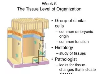
Chapter 4 - Tissue: The Living Fabric • I. Introduction • A. General Characteristics Tissue: group of closely associated cells that are similar in structure and perform a common or related function • 1.) epithelial, • 2.) connective, • 3.) muscle and, • 4.) nervous
Epithelial Muscle Connective Nervous
Functions: • Cover, • Support, • Movement, and • Control
II. Epithelial Tissue` • A. Features • 1. Covers body surface, lines cavities, glandular epithelium • 2. Retain ability to divide throughout life span • 3. 5 functional types of epithelia: • 1. Protective - skin, lining or cavities such as mouth • 2. Exchange - lungs, blood vessels • 3. Transporting - intestine, kidney • 4. Ciliated - nose, trachea • 5. Secretory - sweat and saliva are secreted by epithelia, exocrine glands including pancreas; endocrine including thyroid
B. Special Characteristics • 1. Cellularity: close packed cells • 2. Specialized contacts: tight junctions and desmosomes • 3. Polarity: apical (free) surface and basal surface • 4. Basement membrane: base of the epithelium • a. a network of fine protein filaments embedded in glycoprotein. The filaments hold the epithelial cells to the underlying cell layers - like adhesive junctions • 5. Innervated but avascular - contain no blood vessels. The cells are nourished by diffusion from blood vessels in the underlying connective tissue. • 6. Regeneration - some epithelia are exposed to friction or noxious substances
C. Classification Criteria • a. Cellular shapes: squamous (flat), cuboidal (cube) or columnar • b. Cellular arrangement (layers): simple - 1 cell thick, or stratified - multiple layers • c. Classes • 1) Simple squamous, cuboidal, columnar and pseudostratified • 2) Stratified squamous, cuboidal, columnar and transitional; named for shape of cells at apical surface
Simple Squamous: Single layer of flat cells filtration; kidney lungs, blood vessels 1. Simple Epithelium
single layer as tall as wide secretion and absorption Simple Cuboidal
1) Absorption and, 2) Secretion of mucous Goblet cells secrete mucus Simple ciliated columnar lines uterine tubes and some of respiratory tract Simple Columnar
All cells rest on basement membrane but only tallest reach apical surface Secretion and absorption Pseudostratified ciliated columnar lines most of respiratory tract Pseudostratified Columnar Basement membrane
2. Stratified Epithelia Stratified Squamous: • Most widespread; forms external part of skin and a short distance into body openings • Several layers of cells • So, protective • Surface cells are squamous 5. Deeper cells cuboidal
lines urinary organs(bladder); basal cells cuboidal or columnar 6 layers when empty and 3 layers when stretched Transitional
D. Glandular Epithelia: • General - Secretory epithelia are composed of cells that produce a substance inside the cell and release it - a secretion. • Secretory cells may be scattered or form a gland • a. Gland: one or more cells that make and secrete a particular product or secretion • b. There are 2 types of secretory glands: • endocrine • exocrine
a. Ductless glands b. Release secretions called hormones into blood Endocrine Glands (endo = in)
Exocrine Glands; secrete onto body surfaces Produce 2 secretions: • serous - watery secretions that contain enzymes • mucous - sticky solutions of glycoproteins Release secretions into the external environment; skin surface, epithelial lining. Most use open tubes or ducts. Examples - sweat, mucous, salivary glands
Multicellular Exocrine Glands • a. Contain a duct and secretory unit with secretory cells • b. Structural classification: • 1) Simple glands: single unbranched duct • 2) Compound glands: branched duct • 3) Tubular: secretory cells form tubes • 4) Alveolar: hollow cavity or sac • 5) Tubuloalveolar: both
Connective Tissue • Characterized by the cells widely separated from each other in a matrix that is produced by the cells. • Tissue protects and supports. • Cell Matrix composed of two regions • Ground • Liquid (sol), Gel, Gum or solid • Fibers • Non-elastic (= white or Collagen) • Elastic (= yellow fibers) • Types of Connective tissue
Connective Tissue Types: 1. connective tissue proper ( fat and fibrous tissue of ligaments), 2. cartilage, 3. bone, 4. blood
Regions • ground substance, • fibers, and • cells 1. Ground substance: fills the space between cells and contains fibers 2. Fibers • a. Collagen fibers: primarily collagen; stronger than steel!; white fibers • b. Elastic fibers: long, thin fibers that form networks; stretch and recoil • c. Reticular fibers: fine collagenous fibers continuous with collagen fibers; form networks around vessels, soft tissues and organs and basement membrane
Cells • Each class of connective tissue has a cell type in mature and immature form: • blasts = immature and undifferentiated cells that secrete the ground substance and the matrix • cytes = mature cells
Cells • Cell types in mature and immature form • Fibroblasts: connective tissue proper • Chondroblasts: cartilage • Osteoblasts: bone • Hemocytoblasts: blood • Macrophages: phagocytize foreign materials
blast = bud, forming cyte = less active, mature mode Connective tissues arise from a common origin - mesenchyme
Functions • 1. Binding and structural support • 2. Protection • 3. Insulation • 4. Transportation
Types of Connective Tissue • Loose (Areolar) Connective Tissue • Dense Connective Tissue • Adipose • Cartilage • Bone • Blood
Loose Connective Tissue (Areolar) • Gel like ground with both elastic and non-elastic fibers running though the ground in many directions. • Wraps and cushions organs • Under the skin
Dense Regular Connective Tissue • Nuclei and fibers arranged in parallel rows. • Tendons and ligaments • Fibers mostly non-elastic
Adipose (Fat) • Function as storage cells for adipose (lipids) • Adipose cells contain a large vacuole which in the live cell contains lipids. • Cell nucleus and cytoplasm are pushed out to edge of cell membrane.
Cartilage • Ground of matrix is gum like. • Cells are found in Lacunae within the matrix. • Fibers may be elastic or non-elastic, or a form of non-elastic called reticular(where the non-elastic fibers of very thin) • Hyaline Cartilage-example on the ends of bones • Elastic Cartilage- example ear cartilage • Non-elastic Cartilage- example nose cartilage.
Bone • Ground of matrix is Solid (Calcium carbonate). • Has blood supply and nerves running through the Haversian canal systems.
Vascular Tissue (Blood) • Liquid matrix = plasma • 90% water • 10%Plasma proteins, electrolytes, hormones, oxygen, glucose etc. • Formed elements • Erythrocytes-48billion(female) to 54 billion (male) cell / ml of blood in humans. Mammals are enucleated while rest of the vertebrates they have nuclei • Leukocytes-about 7.5 million / ml of blood • Platelets-blood clotting
Muscle Tissue • Tissue with cells having fibers specialized for contraction. • Skeletal Muscle(Striated, voluntary) • Parallel elongated cells (fibers) • multinucleated and each cell is the length of the muscle. • Light meat, Dark meat—Slow twitch, fast twitch muscle • Smooth Muscle(Visceral, involuntary) • Cells are long and tapered. • Organized into sheets of muscle. • Cardiac Muscle • Intercalated disc • Myogenic • branched
Nervous Tissue • Cells specialized to polarize and depolarize. • Cell is a neuron
Epithelial Membranes • Consists of a least two tissue types: epithelium and connective tissue proper. There are 3 types of membranes • A. Cutaneous • 1. Skin • 2. Keratinized stratified squamous attached to dense irregular connective dermis
B. Mucous Membranes • 1. Line body cavities that open to outside (digestive, respiratory and urogenital) • 2. Mucosae are stratified squamous or simple columnar • 3. Adapted for secretion and absorption • 4. Lamina propria: loose connective tissue under epithelial tissue
C. Serous Membranes (Serosae) • 1. Found in closed ventral body cavities • 2. Parietal and visceral layers, each of simple squamous (mesothelium) on layer of loose connective (areolar) • 3. Serous fluid lubricates between layers • 4. Pleura, pericardium, and peritoneum
VII. Tissue Repair • There are many defenses against damage, but if skin is penetrated, inflammatory and immune responses are activated • A. General • 1. Regeneration: replacement of destroyed tissue with same tissue type • 2. Fibrosis: scar tissue (fibrous connective) forms • B. Inflammation • a. Histamine from mast cells, other chemicals dilate capillaries and make them more permeable • b. Allows WBC's and plasma fluid, rich in clotting proteins and antibodies to enter. • c. Clot forms to stop blood, walls off area, forms a scab. • d. Redness, swelling, heat due to above
2. Organization: First stage of tissue repair; blood clot replaced by granulation tissue (fragile capillaries from nearby that lay down a new capillary bed. • 3. Regeneration and/or fibrosis: epithelium regenerates and scar may remain visible
C. Factors • 1. Tissue type: skin, epithelial, bone, fibrous connective tissues heal well while muscle, cartilage are poor at regeneration • 2. Type of injury and care • 3. Nutrition • 4. Blood supply • 5. Health • 6. Age