Alignment and Structural Analysis of CALTPI and CALTPII Proteins with Tobacco Lipid Transfer Protein 1
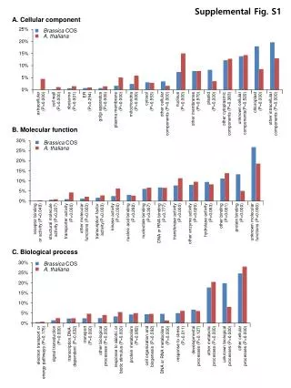
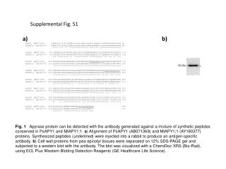
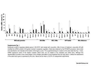
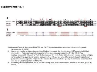
Supplemental Figure 1 illustrates the alignment of CALTPI and CALTPII proteins with the tobacco lipid transfer protein 1 (accession no. D13952). The figure highlights conserved cysteine residues, characteristic of hydrophobic cavity-forming domains in lipid transfer proteins (LTPs), marked with a black diamond (♦). Key features include consensus pentapeptide sequences underlined in red, highly conserved amino acids indicated by arrows, and the predicted secondary structures of CALTPI and CALTPII using Jpred. The alignment was performed using Clustal V in DNASTAR.

Alignment and Structural Analysis of CALTPI and CALTPII Proteins with Tobacco Lipid Transfer Protein 1
E N D
Presentation Transcript
Supplemental Figure 1. Alignment of CALTPI and CALTPII proteins residues with tobacco lipid transfer protein1 (accession no. D13952). • Conserved cysteine residues characteristic of hydrophobic cavity-forming domains of LTPs marked with black diamond (♦). Red underlines indicate position of two consensus pentapeptides, Thr/Ser-X1-X2-Asp-Arg/Lys( residues 63-67) and Pro-Tyr-X-Ile-Ser (residue 101-105) .Red and black arrows indicate position of highly conserved proline, glycine and tyrosine residue involved in loops and turns. Blue arrow indicate the position of putative leader cleavage sit e in lipid transfer proteins. Dashes indicate the spacing in amino acid sequences that was don by ClustV alignment of DNASTAR. • Secondary structure prediction of CALTPI and II using Jpred (http://www.compbio.dundee.ac.uk/~www-jpred/). H, helix E, Supplemental Fig. 1