Chapter 14 Signal-transduction pathways
760 likes | 1.65k Vues
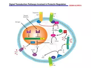
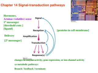
Chapter 14 Signal-transduction pathways. Hormones, Aromas (volatiles) sense 1 messenger [threshold conc.] [ligand]. [proteins in cell membrane]. Delivery [2 messenger]. changes in enzyme activity, gene expression, or ion-channel activity metabolic pathways

Chapter 14 Signal-transduction pathways
E N D
Presentation Transcript
Chapter 14 Signal-transduction pathways Hormones, Aromas (volatiles) sense 1 messenger [threshold conc.] [ligand] [proteins in cell membrane] Delivery [2 messenger] changes in enzyme activity, gene expression, or ion-channel activity metabolic pathways Branch / feedback / terminate
Common second messengers (1) Free to diffuse to other compartments of the cell (2) Significantly amplified signals (3) Common 2º messengers utilized — cross talk: opportunities and potential problems [NO] H2O2 cADP ribose
Signal transduction Signal molecules ligand, primary messenger steroids nonpolar large and polar through membrane bind to receptor bind to protein alter receptor structure intracellular interact with DNA [2° messengers] modulate gene expression protein P / de-P biochemical reaction
Type 1: Seven-transmembrane-helix (7TM) receptor–– serpentine receptor C-terminal and cytoplsmic loop conformation changes rhodopsin ca. 50 % therapeutic drugs
Guanyl nucleotide-binding protein (G protein) an intermediary in signal transduction from 7TM a heterotrimeric G proteins 7TM G-protein-coupled receptors (GPCRs) ATP cAMP
G protein: a heterotrimer : bind the nucleotide (activated and unactivated state) : a seven-bladed propeller : a pair of -helices
(02) G-protein-coupled receptors (GPCRs)
Activated G proteins transmit signals by binding adenylate cyclase ATP cAMP
The epinephrine receptor signal-transduction pathway [cAMP] stimulate ATP production for muscle contraction enhance the degradation of storage fuels increase the secretion of acid by gastric mucosa lead to the dispersion of melanin pigment granules diminish the aggregation of blood platelets induce the opening chloride channel mediated byprotein kinase A(PKA) target protein—ser/thr-P cAMP- response element binding (CREB) protein, in nucleus - a transcriptional activator
Serotonin cAMP- response element binding protein Close K+ channel
How to terminate the signal transduction? 1. Resetting G subunit: a intrinsic GTPase activity,spontaneous seconds ~minutes A build-in clock Mechanism?
Signal termination dep. hormone conc. cytosol Specific (for C-terminal and occupied) diminishes its ability to activate G proteins
Phosphatidyl inositol bisphosphatehydrolysistwo messengers Vasopressin 7TM Gq Phospholipase C Cleavage PIP2 IP3 (soluble form) DAG (insoluble form) A1 A2 D C 1,4,5
DAG and IP3 work in tandem
(02) IP3 open channels to release Ca2+ 3IP3 + IP3 -gate channel Ca2+ release (from ER, SR) Smooth muscle contraction Glycogen breakdown Vesicle release Early fertilization (nM) a short-lived messenger
Diacylglycerol metabolism (02) phosphorylated Arachidonate Prostaglandin H2 hydrolyzed
Ca2+— a ubiquitous cytosolic messenger Plagiarism The reasons for Ca2+ mediate many signaling processes 1. Fleeting changes in [Ca2+] are readily detected The low level of [Ca2+]cyto can be easily and abruptly raised for signaling purposes100 nM avoid insoluble compound formation (an apparent drawback is in fact an advantage) is via Ca2+-ATPase and Na+- Ca2+ exchanger (02) 2. Ca2+ can bind tightly to proteins and result in significant conformational changes coordinated with six to eight oxygen atoms from proteins or water 6 O from aa/protein 1 O from water
How to detect or monitor the variation of [Ca2+] in real time - The fluorescent calcium-binding dye Red: high Blue: low
Calcium-specific reagents (02) ionophores raise the cytosolic Ca2+ level reduce the unbound Ca2+ level EDTA: ethylenediamine tetraacetic acid
Calmodium – a calcium sensor ¤ a 17-kd with 4 calcium-binding sites ¤ is activated when [Ca2+]cyto 500 nm ¤ a member of EF-hand protein family, a calcium binding motif - a helix-loop-helix unit ¤ Parvalbumin: vitamin D3-dependent Ca2+ binding proteins forefinger thumb
Calmoduline-dependent protein kinase (CaM kinase)– recognize positively charged, amphipatic helix Calcium bind calmodium conformational changes expose hydrophobic surfaces that can be used to bind other proteins A pair of EF-hand motifs a flexible helix
(02) calcium + calmodulin CaM kinase Ca2+-ATPase pump active target protein [Ca2+]cyto level decrease signal propagation signal termination (the memory of a previous calcium pulse)
Type 2: Insulin signaling receptors that include protein kinase as part of their structures receptor is a dimer of two identical units each unit: - and -chain linked by a disulfide bond -chain -chain One insulin binding on the outside of the cell A membrane-associated kinase within the cell is activated; cross-phosphorylation 2 inter- and 1-intra-chain disulfide bond
Protein kinase A: Ser/Thr P in subunit 3 tyr residues in activation loop of subunit
a series of membrane-anchored molecules IRS : insulin-receptor substrate
IRS : insulin-receptor substrate IRS1/IRS2, act as adaptor proteins N-terminal: Pleckstrin homology domain, binds phosphoinositide lipids phosphotyrosine-binding domain: Tyr-X-X-M sequence: are phosphorylated by the receptor tyrosine kinase Met
IRS phosphoinositide 3-kinase: a lipid kinase, 110 kd catalytic subunit and 85 kd regulatory subunit containing aSH2 domain: Src homology 2, recognize the phosphotyrosine residues in the IRS, via two Arg residues that are conserved in all SH2 domain
phosphoinositide 3-kinase PIP3 PIP3-dependent protein kinase Akt: a kind of protein kinase, is not membrane anchored
Membrane-anchor molecules Glucose transporters (GLUT4) Stimulate glycogen synthesis
Amplicification/ termination phosphatase phosphatase phosphatase
Type 3: EGF (epidermal growth factor) signaling stimulate the growth of epidermal and epithelial cells a receptor tyrosine kinase, a 6 kd polypeptide 3 intrachain disulfide bonds EGF receptor structure
EGF receptor: is a dimer of two identical units, but exist as monomers until EGF ligands bind to them each monomer binds a EGF molecule in its extracellular domain each EGF molecule lies far away from the dimer interface a dimerization arm from each monomer that reaches out and inserts into a binding pocket on the other monomer
If EGF is absent? binds to a part of within the same monomer Once EGF present, Change into a active conformation A constitutive active form ? Her 2 receptor, 50 % identical in aa sequence with the EGF receptor and has the same domain structure Her 2 is overexpressed in some cancers
EGF phosphorylation: also like insulin receptor, cross-phosphorylation of one unit by another unit within a dimer, but its carboxyl - terminal tail containing tyrosine rich (5 residues) the kinase itself is an active conformation without phosphorylation Dimerization C-terminal region on one receptor into the active site of its partner’s kinase
Grb-2: an adaptor protein SH2 domain phosphotyrosine residues of receptor SH3 domain proline-rich region of Sos
Sos: a guanine-nucleotide-exchange factor (GEF) Ras: small G proteins, small GTPase localized to the inner surface of plasmamembrane
two GTPase-activating proteins (GAPs) phosphatase
G proteins vs. small G proteins(divergent evolution) G proteins small G proteins 30-35 kd 20-25 kd heterotrimer monomer (similar to G) 7TM dimerization GTPase act. GTPase act. (low) GTPase-activating proteins (GAPs): facilitate GTP hydrolysis Sos + GAPs adjust small G cycle ras mutation cancer
14.4Many elements recur with variation in different signal transduction pathways Protein kinases are central Second messengers Specialized domains pleckstrin homology domains: interact with lipids PIP3 SH2 domains: interact with the phosphorylated tyrosine residues
Some virus induced cancer– to understand the signal-transduction proteins and pathways Rous sarcoma virus: a retrovirus, a oncogenic RNA virus viral sarcoma (v-src): oncogene[A cancer-causing gene; any of several mutant genes that cause cells to exhibit rapid, uncontrol proliferation.] cellular sarcoma (c-src): proto-oncogene, does not induce cell transformation v-Src: 11 aa of C-terminal, lack Y residue always active c-Src 19 aa a. SH2 bind to tyr-P of C-terminal b. The linker between SH2 and protein kinaseis bounded by SH3 c-Src inactive Biology/chemical/physical factors
Ras: a small G protein or GTPase– localized to the inner surface of plasmamembrane The small G proteins Three 21-kd Ras proteins in mammalian cells H-Ras: Harvey rat sarcoma K-Ras: Kirsten rat sarcoma A loss of the ability to hydrolyze GTP N-Ras: Neuroblastoma rat sarcoma continue on Tumor-suppressor genes (contribute to cancer development): to develop cancer only when both copies of the genes normally present in a cell are deleted or otherwise damaged. e.g., genes for some of the phosphatase
Monoclonal antibodies utilization: inhibit the signal transduction in activated tumor formation In some human epithelial cancers, such as breast, ovarian, and colorectal cancers, overexpressed the epidermal-growth-factor receptor (EGFR) Monoclonal Ab offend receptor e.g., Cetuximab, target a receptor tyrosine kinase Trastuzumab (Herceptin): inhibit Her2 overexpressed in breast cancers
Protein kinase inhibitor– a potential anticancer drugs Chronic myologenous leukemia (CML) chromosome defect: the translocation between chromosome 9 and 22 (reciprocally) Bcr-Abl fused protein: overexpress kinase activity and is not regulated appropriately STI-571: a specific Bcr-Abl kinase inhibitor encode tyrosine kinase To understanding the signal-transduction pathways is leading to conceptually new disease treatment. Metabolism disease
Choleragen a cholera toxin from Vibrio cholera (G -) two functional units: subunit B: bind to GM1 gangliosides of intestinal epithelium (p. 738) subunit A: enters the cell, catalyze the covalent modification of Gs protein Gs + subunit A Gs-Arg-ADP-ribose stabilize Gs-GTP form (perpetually stimulation) activate adenylate cyclase [cAMP] activate protein kinase A open Cl- channel / inhibit Na+-H+ exchanger NaCl and H2O loss Treatment consists of rehydration with a glucose-electrolyte solution.