The cytoskeleton The cell surface and junctions
390 likes | 560 Vues
The cytoskeleton The cell surface and junctions. Cytoskeleton. The cytoskeleton is the structure consisting of fibrous proteins that occur in the cytoplasm and maintain the shape of the cell. Cytoplasm.

The cytoskeleton The cell surface and junctions
E N D
Presentation Transcript
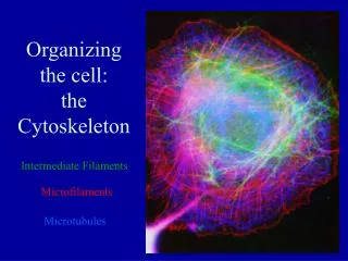
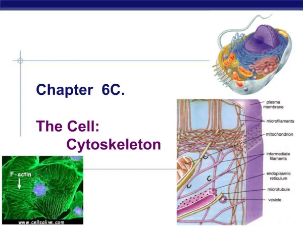
Cytoskeleton The cytoskeleton is the structure consisting of fibrous proteins that occur in the cytoplasm and maintain the shape of the cell.
Cytoplasm Microtubules – function in cell division and serve as a "temporary scaffolding" for other organelles. Actinmicrofilaments are thin threads that function in cell division and cell motility. Intermediate filaments are between the size of the microtubules and the actin filaments.
The Cytosceleton • The cytoskeleton: • gives the cell shape, • anchors some organelles and • directs the movement of others, • and may enable the entire cell to change shape or move.
The Cytosceleton It may play a regulatory role, by mechanically transmitting signals from the cell's surface to its interior.
Role of microtubules • Hollow tubes with wall that consists of 13 columns of tubulin molecules (25 nm in diameter) • Involved in: • cell shape maintenance (compression resistance) • cell motility (as in cilia or flagella) • chromosome movement in cell division Organelle movements
Motor molecules and the cytoskeleton The microtubules and microfilaments interact with proteins called motor molecules. Motor molecules change their shapes, moving back and forth something like microscopic legs. ATP powers these conformational changes.
Motor molecules and the cytoskeleton • The motor molecule releases at its free end and then grips at a site further along a microtubule or microfilament. • For example, a sliding of neighboring microtubules moves cilia and flagella.
Motor molecules and the cytoskeleton In muscle cell contraction, motor molecules slide microfilaments rather than microtubules. (b) Motor molecules can also attach to receptors on organelles such as vesicles and enable the organelles to "walk" along microtubules of the cytoskeleton.
Motor molecules and the cytoskeleton For example, vesicles containing neurotransmitters migrate to the tips of axons, the long extensions of nerve cells that release transmitter molecules as chemical signals to adjacent nerve cells.
Motor molecules and the cytoskeleton Kinesin moves organelles towards periphery (+), Dinein towards the nucleus (-).
Centrosome containing a pair of centrioles An animal cell has a pair of centrioles within its centrosome, the region near the nucleus where the cell's microtubules are initiated. The centrioles, each about 250 nm (0.25 μm) in diameter, are arranged at right angles to each other, and each is made up of nine sets of three microtubules (TEM).
Flagella and Cilia Locomotive appendages that protrude from some cells. A specialized arrangement of microtubules responsible for their beating
A comparison of the beating of flagella and cilia (a) A flagellum has a snakelike motion driving a cell in the same direction as the axis of the flagellum. Propulsion of a sperm cell is an example of flagellate locomotion (SEM).
A comparison of the beating of flagella and cilia (b) The cilia of Paramecium beat at a rate of about 40 to 60 strokes per second. Cilia have a back-and-forth motion, alternating active strokes with recovery strokes. This moves the cell, or moves a fluid over the surface of a stationary cell.
The 9+2 arrangement of microtubules in a flagellum or cilium.
Ultrastructure The basal body anchoring the cilium or flagellum to the cell has a ring of nine microtubule triplets. The nine doublets of the cilium extend into the basal body, where each doublet joins another microtubule to form the ring of nine triplets. The two central microtubules of the cilium terminate above the basal body (TEM).
Dynein – motor protein Responsible for the bending movements of cilia and flagella The dynein arms of one microtubule doublet grip the adjacent doublet, pull, release, and then grip again. The action of the dynein arms causes the doublets to bend.
Actin and tubulin components of the cytoskeleton. Microfilaments – actin filaments. They are built from molecules of a globular protein – actin. A microfilament is a twisted double chain of actin subunits (7 nm in diameter)
Role of microfilaments Maintenance of cell shape (as a tension-bearing elements) Changes in cell shape Muscle contraction Cytoplasmic streaming Cell motility Cell division – cleavage furrow formation
A structural role of microfilaments The surface area intestinal cell is increased by its many microvilli, cellular extensions reinforced by bundles of microfilaments. These actin filaments are anchored to a network of intermediate filaments
Microfilaments and motility (a) In muscle cells, actin filaments (orange) lie parallel to thick myosin filaments (purple). Myosin acts as a motor molecule. The teamwork of many such sliding filaments enables the entire muscle cell to shorten.
Microfilaments and motility (b) In a crawling cell (ameboid movement), actin is organized into a network in the gel-like cortex (outer layer). This contraction forces the interior fluid into the pseudopod, where the actin network has been weakened. The pseudopod extends until the actin reassembles into a network.
Microfilaments and motility (c) In cytoplasmic streaming, a layer of cytoplasm cycles around the cell, moving over a carpet of parallel actin filaments. Myosin motors attached to organelles in the fluid cytosol may drive the streaming by interacting with the actin.
Role of intermediate filaments Fibrous proteins supercoiled into thicker cables (8-12 nm) Depending on the cell type, it is presented by one of the several different proteins of the keratin family • Responsible for: • maintenance of cell shape (tension-bearing elements) • anchorage of nucleus and certain other organelles • formation of nuclear lamina
Plant cell walls • The wall: • Protects the plant cell, • Maintains its shape, • Prevents excessive uptake of water • Holds the plant up against the force of gravity
Plant cell walls Young cells first construct thin primary walls. Stronger secondary walls are added to the inside of the primary wall when growth ceases. A sticky middle lamella cements adjacent cells together.
Plant cell walls • The walls do not isolate the cells: • the cytoplasm of one cell is continuous with the cytoplasm of its neighbors via plasmodesmata, channels through the walls (TEM).
Extracellular matrix (ECM) of an animal cell Glycoprotein collagen fibers are embedded in a web of proteoglycans, which can be as much as 95% carbohydrate; the proteoglycan molecules have formed complexes by noncovalently attaching to long polysaccharide molecules.
Extracellular matrix (ECM) of an animal cell The third glycoprotein is fibronectin, the adhesive that attaches the ECM to the plasma membrane of the cell. Membrane proteins called integrins are bound to the ECM on one side and to the microfilaments of the cytoskeleton on the other. This linkage can transmit stimuli between the cell's external environment and its interior.
Intracellular junctions in animals (a) Tight junctions - continuous belts around the cell. Fusion of neighboring cell membranes forms a seal that prevents leakage of extracellular fluid across a layer of epithelial cells.
Intracellular junctions in animals The tight junctions of the intestinal epithelium keep the contents of the intestine separate from the body fluid on the opposite side of the epithelium.
Intracellular junctions in animals (b) Desmosomes (anchoring junctions) fasten cells together into strong epithelial sheets. Intermediate filaments made of the protein keratin reinforce desmosomes (TEM).
Intracellular junctions in animals (c) Gap junctions (communicating junctions) provide cytoplasmic channels between adjacent cells. Special membrane proteins surround each pore. The pore is wide enough for salts, sugars, amino acids, and other small molecules to pass (TEM).
Intracellular junctions in animals In the muscle tissue of the heart, the flow of ions through gap junctions coordinates the contractions of the cells. Gap junctions are especially common in animal embryos, in which chemical communication between cells is essential for development.
Reading Campbell et al. Biology. Ch. 6 A tour of the cell, 112-124