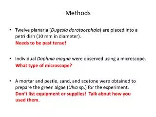
Methods
Comparative In Vitro Toxicity Gabrielle Maldonado 1,2 , Mark Wilson 1,3 1 Emerging Scholars Environmental Health Science Academy, 2 Chalmette High School 3 Department of Global Environmental Health Sciences, Tulane University School of Public Health and Tropical Medicine. Abstract.

Methods
E N D
Presentation Transcript
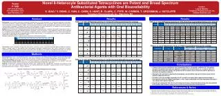
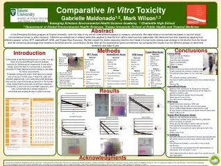
Comparative In Vitro Toxicity Gabrielle Maldonado1,2, Mark Wilson1,3 1 Emerging Scholars Environmental Health Science Academy, 2 Chalmette High School 3 Department of Global Environmental Health Sciences, Tulane University School of Public Health and Tropical Medicine Abstract In the Emerging Scholars program at Tulane University, I with the help of my mentor used different assays to measure cytotoxicity. We used ethanol concentrations based on alcohol blood concentrations known to affect humans. Different concentrations of ethanol were then applied to Hep G2 liver cells to see how they responded. We observed how they reacted by applying five different assays: colony, MTT, alamarBlue®, SRB, and Trypan Blue Exclusion. We then raised the doses reasoning that the liver inside a human body cleans a percentage of the alcohol from the blood and the remaining percentage that remains is the blood alcohol concentration levels. After all the experiments were commenced, we compared the results from the different assays to determine sensitivity and ease of use. Conclusions Methods Introduction Trypan Blue Assay • Colony Assay • Based on cell proliferation • Uses petri dishes • Uses the most materials • Easy to contaminate • Takes ≈ 2 weeks • MTT Assay • Based on working mitochondrial functions • Purple crystals are formed in healthy cells • The cells are then lysed, so it’s a one way • trip • The crystals undergo solubilization • The more healthy cells there are the more purple the dye gets • Takes a day • alamarBlue® Assay • Based on the conversion of resazurin to resorufin • Resazurinbecomes fluorescent due to the reductive reactions of active cells • More fluorescence equals more living cells • Takes about 5 hours to do • The cells can be used again • SRB Assay • Based on protein binding of SRB dye • The binding fixes the cells onto the well plates • Doesn’t differentiate between dead and live cells • At high doses of ethanol can fix dead cells and skew data • Takes 3 days to do • Trypan Blue Exclusion Assay • Based on cell membrane structure • Trypan blue is added and is absorbed by dead cells • A lot of counting is required • There are several steps in which cells can be lost • It isn’t practical for continuous lab work • The data isn’t very good as many cells were lost in the process • Takes a day to do • In Summary: • alamarBlue® was the least time intensive and agreed well with other metrics of cytotoxicity, and the cells were still viable. • The difference between in vivo and in vitro is worth mentioning. • - In vitro testing is not always the best representation of • what goes on in the body (in vivo) • Other testing should be done to see if our results are cell type dependent • Colony Assay • Materials • Petri dishes • Medium • Ethanol • Hep G2 cells • Crystal Violet Method • Set up petri dishes • Treat with ethanol • Change the medium • Put in incubator and wait ≈ 2 weeks • Take off medium • Wash with PBS • Stain with crystal violet • Count cell colonies • MTT Assay • Materials • 96 well plates • Medium • HepG2 cells • Ethanol • MTT • Method • Set up 96 well plate • Treat with ethanol • Add MTT • Incubate for 2-4 hours • Put in spectrophotometer • And visualize results: More cells= more purple • alamarBlue® Assay • Materials • 96 well plates • Medium • HepG2 cells • Ethanol • alamarBlue® • Method • Set up 96 well plate • Treat with ethanol • Add dye • Put in fluorescence scanner: • excitation at 548 & emission • at 585 SRB Assay • Materials • 96 well plates • Medium • Hep G2 cells • Ethanol • centrifuge • Trypan Blue • Method • Set up 96 well plate • Treat the cells with ethanol • Trypsinize to get cells off plate • Stain w/ trypan blue • Put in centrifuge • Count w/ hemocytometer • Materials • 96 well plates • Medium • Hep G2 cell • Ethanol • SRB • Method • Set up 96 well plate • Treat with ethanol • Fix cells • Stain cells • Wash cells • Read plate Cytotoxicity is defined as being toxic to cells. In a lab there are several different ways to assess cytotoxicity. Setting up an experiment to see which assay works the best for cytotoxicity is important because it’ll help determine which assay to use later for future experiments. To assess cytotoxicity, there first has to be a target and a toxicant. In this case, it was liver cells and ethanol respectively. Liver cells were used because they are the ones that break up alcohol in the human body. Experiments were set up with two exposure time points, a one hour and overnight exposure, and with concentrations of ethanol based on concentrations already known to affect humans. Results • A hybridizing of effects as described at Alcohol's Effects from Virginia TechandFederal Aviation Regulation (CFR) 91.17: Alcohol and Flying (hosted onFlightPhysical.com) • BAC Charts from Virginia Tech Acknowledgments I would like to thank Dr. Wickliffe for the use of his lab. I would also like to thank Mrs. Perrault for bringing me into this program. I thank my family for supporting me as always. Finally, I thank the Ph. D students on the 21st floor for giving me nudges in the right direction. This work was supported by the Gulf Region Outreach Program (GRHOP) which is funded from the Deepwater Horizon Medical Benefits Class Action Settlement approved by the U.S. District Court in New Orleans on January 11, 2013.