Understanding Immune Recognition
540 likes | 721 Vues
Understanding Immune Recognition. B cells can recognise antigens via their surface Ig molecules T cells can only recognise antigen in association with a Major Histocompatibility Complex (MHC) molecule. Antigen Recognition. B cells can recognise antigens via their surface Ig molecules

Understanding Immune Recognition
E N D
Presentation Transcript
B cells can recognise antigens via their surface Ig molecules T cells can only recognise antigen in association with a Major Histocompatibility Complex (MHC) molecule. Antigen Recognition
B cells can recognise antigens via their surface Ig molecules T cells can only recognise antigen in association with a Major Histocompatibility Complex (MHC) molecule. Antigen Recognition
Immunoglobin Fold • V and C domains share the basic Ig fold • Differences between the two domains • C domain is built of seven b-strands arranged so that four strands form one sheet and three strands form a second sheet. • The strands are closely packed together and joined by a single disulphide bond • Most of the invariant residues of the constant domain are in the sheets • Overall structure of the V domain very similar but there are nine strands instead of seven. The two additional strands harbour CDR2
The Complementarity Determining Regions • Six loops of the VH (H1, H2 and H3) and VL (L1, L2 and L3) domains create a great variety of surfaces • Deep binding cavities: such as those seen in some antibody-hapten complexes • Wide pockets : seen in certain antibody-peptide complexes • Flat surfaces : seen in antibody-protein interactions • H3 is the most variable of the loops and in all crystallographically solved antibody-antigen complexes makes several contacts with antigen
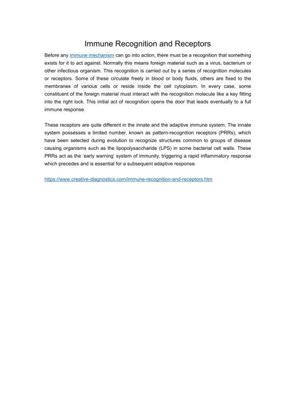
Proteins (conformational determinants, denatured or proteolyzed determinants) Nucleic acids Polysaccharides Some lipids Small chemicals (haptens) What Do Antibodies Recognize?
Antigen:Antibody complex • Antibodies bind to antigens by recognizing a large surface, and through surface complementarity. • Thus, these complexes have a very high affinity for each other.
Weak forces vs high affinity • The interaction between an antigen and antibody can be very strong, and yet all of the forces involved are considered to be relatively weak. How can weak hydrogen bonds, electrostatic attractions, hydrophobic forces, and van der Waals contacts lead to a high affinity? • Contact between antigen and antibody occurs over a wide surface area, allowing multiple weak interactions that give a strong affinity • Hydrogen bonds join the antibody and antigen over a wide surface area. Other weak forces, including van der Waals forces, electrostatic attractions and hydrophobic forces, add to the strength and specificity of antibody/antigen binding
Antibody-Hapten Complex • Haptens, having a limited total surface area, deeply embed themselves into the VL/VH dimer interface • Hapten binding antibodies frequently show a deep central cavity, long CDR L1 loops and a CDR H3 loop with an "open" conformation, allowing the hapten to bind as much as 80% of its total surface in the interaction.
Protein Antibody Complex • In contrast, proteins preferentially to a relatively flat binding surface • In a "closed" CDR H3 conformation, the CDR H3 loop packs down onto the central cavity, and the protein antigen binds on top of it.
Antibodies not only must recognize antigen, but also must invoke responses – effector functions – that will remove the antigen and kill the pathogen. Variable regions of antibody are the sole agents of binding to antigen. The heavy chain constant region (CH) is responsible for interactions with other proteins (e.g. complement), cells (elements of innate immune system), and tissues that result in the effector functions of the humoral response. FcR recognize the Fc portion of antibodies not antigens Effector response is mediated via Ig-FcR complex formation
The Fc-Fc Receptor complex • FcR plays important role in antibody mediated immune responses • Ig and FcR binding activates effector functions • Fc Receptor interacts with the CH2 and CH3 domains of Immunoglobulins
B cells can recognise antigens via their surface Ig molecules T cells can only recognise antigen in association with a Major Histocompatibility Complex (MHC) molecule. Antigen Recognition
T cells display TCR as their antigen recognition protein When stimulated they become Cytotoxic or Helper T cells Secrete cytokines that recruit other cells of the IS TCR’s only recognise short peptides. T cells
T cells have a requirement to recognise both the ANTIGEN and the MHC molecule. This is because the molecular structure of the MHC-Antigen complex is arranged so that some of the polymorphic amino acids of the MHC molecule are in direct contact with the TCR Therefore T cell recognition of antigen is said to be MHC ‘restricted’. MHC & T cells
Fragmentation of protein into peptides Association of peptide with an MHC molecule Transport to cell surface for expression Different cellular pathways for association of peptide with MHC class I and class II molecules Antigen Processing and Presentation
MHC Class I present endogenously derived peptides. these can be either self or derived from viruses because MHC Class I is present on all cells any cell can interact with T cells if infected by a virus MHC Class II present exogenous antigen which has been phagocytosed and processed.eg. Bacteria This is performed by professional antigen presenting cells eg macrophages MHC & Antigens
MHC Class I detected on all nucleated cells very highly polymorphic Tight fit for peptides of only about 9 aa consists of an a-chain of 3 domains associated with b-2 microglobulin MHC Class II seen only on the ‘professional antigen processing cells’ e.g macrophage slightly less polymorphic accepts peptides of up to 15 aa acids MHC
CD8 T-CELL CD4 T-CELL CD3 b CD3 a a b a b CD8 TCR ab TCR ab CD4 9 aa peptide 15 aa peptide a2 a1 a1 b1 MHC b2m MHC CLASS I CLASS II a3 b2 a2 ANTIGEN PRESENTING CELL ANTIGEN PRESENTING CELL MOLECULES OF T LYMPHOCYTE RECOGNITION • Major histocompatibility complex (MHC); human=Human Leukocyte Antigen (HLA); mouse=H-2 • Gorer and Snell identified a genetic basis for graft rejection and Snell named it histocompatibility 2 (H-2). Nobel prize awarded to Snell. • Highly polymorphic genes organized in a complex on chromosome 6 (human) and 17 (mouse). • Glycoproteins expressed on the surface of cells. MHC class I is composed of one polypeptide, non-covalently associated with b2microglobulin. MHC class II is composed of two polypeptides, referred to as a and b.
Class I Alpha Chain 3 External domains 1 Transmembrane 1 Cytoplasmic tail Encoded in MHC Beta-2 Microglobulin 1 External domain No transmembrane No Cytoplasmic tail Not encoded in MHC Class II Alpha Chain 2 External domains 1 Transmembrane 1 Cytoplasmic Tail Encoded in MHC Beta Chain 2 External domains 1 Transmembrane 1 Cytoplasmic Tail Encoded in MHC MHC Class I and Class II Proteins
Peptides bind to MHC molecules in a polyproline II conformation
Peptides of intracellular origin Peptides 9-10 residues long Deep pockets bind peptide sidechains Deep pockets bind peptide N- and C-termini Peptides of extracellular origin Peptides 15 residues or longer Shallow pockets bind peptide sidechains Peptide termini free H-bonds to peptide backbone Peptide Binding by Major Histocompatibility Complex (MHC) Antigen-presenting Proteins MHC I MHC II
Both Class I and Class II genes are highly polymorphic Most polymorphic residues of Class I are in the alpha 1 and alpha 2 domains Most polymorphic residues of Class II are in the alpha 1 and beta 1 domains MHC Polymorphism
Allelic variation in MHC occurs at the peptide binding site and on the top/sides of the binding cleft
The T cell receptor (TCR) is a complex of integral membrane proteins that participates in the activation of T cells in response to the presentation of antigen. Specific recognition and binding by the clonotype-specific a/b heterodimer leads to activation of transcription and commitment of the T cell to CD4+ or CD8+ fate. This activation involves other subunits of the receptor complex as well as other membrane-associated molecules that couple the extracellular liganding event to downstream signaling pathways such as protein phosphorylation, the release of inositol phosphates and the elevation of intracellular calcium levels.
TCR binds peptide/MHC with a restricted (but variable) orientation
peptide binding interface: 21-34% proportion of TCR contacts with the peptide:26-47% contact are different between TCR-MHC complex -the contribution to the binding energy is still uncleared! Bandovich and Garcia. 2003. Immunity 18,7-11
TRI-MOLECULAR COMPLEX CHARACTERISTICS -CDR1 and CDR2 interact with MHC molecules (a helices) -CDR3 interacts with the peptide -interaction always in the same orientation -45 to 70 degrees angle related to peptide -Va see N-ter of the peptide -Vb see C-ter of the peptide
TRI-MOLECULAR COMPLEX CHARACTERISTICS - most of the binding interface is between the TCR and MHC helices - conformational change in the TCR CDR loops enhances TCR crossreactivity - no conformational change in the TCR constant region (except in one complex out of ten)