Download
1 / 36
360 likes | 454 Vues
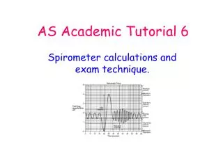
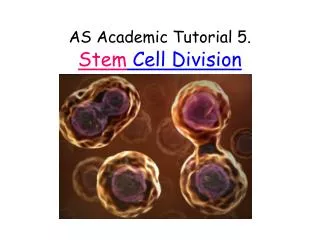
Explore the process of stem cell division, from the replication of DNA to cytokinesis, and understand the role of chromosomes. Learn about the stages of the cell cycle and mitosis, including interphase, prophase, metaphase, anaphase, telophase, and cytokinesis. Delve into the significance of protein synthesis, organelle synthesis, and DNA replication in the cell cycle. Enhance your knowledge with practical activities like mitotic phase identification and biological drawings. Prepare for quizzes and exam questions to test your understanding of stem cell division.

E N D
So what are chromosomes? • http://advancedsciences.cambridge.org/ocr/biology_1/animation/878
Let’s revise the cell cycle Cytokinesis: cytoplasm divides Nucleus divides growth new protein and organelles made mitosis G1 Proteins needed for cell division made G2 S DNA replicates
True or False? • The longest stage in the lifecycle of a stem cell is mitosis. • Mitosis is the division of a nucleus. • Replication of the DNA to produce identical sister chromatids occurs in the G2 phase. • The products of the cell cycle are genetically identical cells. • If there are 10 units of DNA in the original cell in G1, there are 20 in each of the new cells after cytokinesis.
So, before mitosis occurs……. Cell at interphase • Protein synthesis • Organelle synthesis • DNA replication
A single chromosome at the beginning of mitosis Has two chromatids, as the DNA replicates during interphasebefore mitosis centromere
3 happenings occur in prophase. Early prophase centrioles nucleolus DNA coils and condenses Chromosomes take up stain
prophase Centrioles move apart Nuclear membrane breaks down Nucleolus fades Chromosomes continue to condense
Nucleolus disappears Chromosomes continue to condense What were the 3 happenings?
metaphase Chromosomes aligned on the equator Nuclear membrane broken down Each chromosome attached to spindle fibre by the centromere
Anaphase Centromeres divide and chromatids pulled apart
Anaphase Sister chromatids are pulled to opposite poles of the cell
Mitosis Animations! • http://uk.youtube.com/watch?v=nPG6480RQo0&feature=related • Musical introduction! • http://uk.youtube.com/watch?v=ELVwj5JDfg8&feature=related • http://www.youtube.com/watch?feature=iv&src_vid=nPG6480RQo0&v=VGV3fv-uZYI&annotation_id=annotation_706798 3D good • http://www.youtube.com/watch?v=cvlpmmvB_m4&feature=endscreen&NR=1 Very clear - good • http://www.cellsalive.com/mitosis.htm
In a moment you will be preparing a root tip squash to look for stained cells like these. Let’s recognise the phases! Interphase
So now to the practical – see instructions in booklet page 4……Draw any 2 cells you find showing different mitotic phases, or 2 cells from the prepared slides. • Biological Drawings • Use the whole page. • Sharp pencil • No shading • No sketching • Labels with arrow heads.
When you have finished…….. • Tidy all your equipment carefully away. • Start answering the quizzes in the booklet pages 1 – 3 and 5, and then go on to the exam questions pages 8 - 13. They start easy and become challenging! • Page 8 should read “ Fig 5.1 shows drawings of nuclei, A to D, from 2 different plant species. Two of them show prophase of mitosis.”