Download
1 / 32
320 likes | 474 Vues
Shoulder Reconstruction. Department of Orthopaedic, CKUH Sen-Jen Lee Reference: Orthopaedic Knowledge Update 6. Muscular Function and Anatomy of the Glenohumeral Joint. Static stabilizer: Capsuloligamentous structures Superior, middle, and inferior GH ligaments Dynamic stabilizer:

E N D
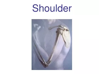
Shoulder Reconstruction Department of Orthopaedic, CKUH Sen-Jen Lee Reference: Orthopaedic Knowledge Update 6
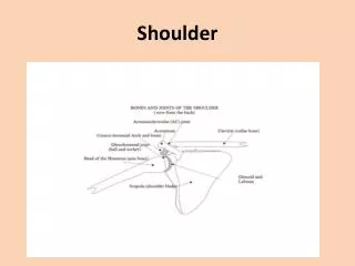
Muscular Function and Anatomy of the Glenohumeral Joint • Static stabilizer: • Capsuloligamentous structures • Superior, middle, and inferior GH ligaments • Dynamic stabilizer: • Rotator cuff muscles • Center the humeral head in the glenoid fossa • Long head of the biceps tendon • Proprioceptive mechanisms • Ruffini receptors and pacinian corpuscles • Ligamentomuscular reflex arcs
Arthroscopy of the Shoulder • As a diagnostic tool • Arthroscopic subacromial decompression • For treating frozen shoulder and rotator cuff tears • For treating superior labral tears (SLAP lesions) • For treating dislocating or subluxating shoulders
Rotator Cuff Disease • Etiology • Mechanical impingement • Compression of the supraspinatus tendon between the acromion and the greater tuberosity • Intrinsic degenerative processes within the aging tendon • Tendon inflammation, tendon and bursal fibrosis, tendon tears (partial- or full-thickness), and cuff tear arthropathy • Acromial morphology • Flat, curved, or hooked
Rotator Cuff Disease • In a biomechanical study • The acromial undersurface and rotator cuff were in closest proximity between 60° and 120° of elevation • Contact was consistently centered on the supraspinatus insertion • Intrinsic histological and mechanical properties • The bursal-side layer: tendon bundles • The joint-side layer: a complex of tendon, ligament, and joint capsule • The strain-to-yield point and ultimate failure stress • Bursal-side layer were twice as great as those of the joint-side layer
Impingement Syndrome • Common cause of shoulder pain • Clinical diagnosis • History and physical examination • Radiographs • Supraspinatus outlet view: • Subacromial spurs and the morphology of the acromion • Functional impingement instability • Internal impingement • Impingement of the undersurface of the rotator cuff on the posterior glenoid rim
Pathogenesis of Rotator Cuff Lesion Overuse 10 Impingement -Outlet Stenosis 20 Impingement -Instability 10 Degeneration -Insubstance tears -Aging -Avascularity Extrinsic Intrinsic Rotator Cuff Injuries Tendinitis / Tendinosis
Three Stages of Impingement Lesions • Stage I: edema and hemorrhage • Reversible lesion, < 25 years old • Stage II: fibrosis and tendinitis • Recurrent pain with activity, 25 - 40 years old. • Stage III: tears of the rotator cuff, biceps ruptures, and bone changes • Progressive disability, > 40 years old. • Neer C.S ii, 1983
Extrinsic Factors • 95 % of RCT are initiated by impingement wear rather than circulatory impairment or trauma. • Shape and slope of the acromion. • Impingement wear, then “acute extension” of a tear. • Neer II, JBJS,1972 & Cli.Orthop, 1983
Intrinsic Factors • Partial articular-sided tears with normal acromial morphology • Cuff degeneration (aging and trauma) RCT • Ozaki et al: JBJS, 1988 (A study in cadaver) • Inflammation Angioblastic hyperplasia fibrosis, calcification, RCT. • Nirschl et al: Instr. Course Lect. 1989
Diagnosis of Impingement Syndrome: Hx, PE RCT: sensitivity:91% specificity: 75 % Ext. R Int. R. Elevation
Neer imp.sign Hawkin imp. sign Painful arc Imping. Test Supraspinatus test
Lift-off test Speed’s test
Image Study of the Rotator Cuff • X-ray: scapular AP/Lat • Arthrogram • MRI • Ultrasonogram
Ultrasonogram of the Shoulder(I) • High resolution, real-time equipment • A 7.5 MHz linear array transducer • ATL’s high definition imaging (HDI) 5000 (NCKU)
Impingement Syndrome Nonsurgical Treatment • Corticosteroid injections • Better pain relief and greater increases in active motion • No more than 2 subacromial cortisone injections • Be avoided in patients with rotator cuff tear • Anti-inflammatory medications and physical therapy • 67% satisfactory results
Impingement Syndrome Surgical Treatment • Open acromioplasty • More excellent results • Arthroscopic acromioplasty • Reduced early perioperative morbidity • Easier rehabilitation • Decreased hospitalization time • Ability to detect and treat concomitant glenohumeral pathology • Better preservation of the deltoid origin • A smaller surgical scar
Impingement Syndrome • Failure of arthroscopic acromioplasty • Improper diagnosis • Inadequate bone removal • Technical errors • Overaggressive bone removal leading to deltoid injury or in rare cases to acromial fracture
Partial-thickness Tears • Partial-thickness tears • Magnetic resonance imaging (MRI) and arthroscopy • Arthroscopic debridement and acromioplasty • Recent study: (>50% thickness of the tendon ) • 15/32 good results in arthroscopic debridement and acromioplasty • 31/33 excellent or good results in arthroscopic acromioplasty and mini-open repair
Full-thickness Tears • Symptomatic full-thickness rotator cuff tears • Anterior acromioplasty and rotator cuff repair • Factors in decision-making • Severity and duration of symptoms • Functional limitations • Patient demands and expectations • Tear size, and tear location • Factors affect the results of rotator cuff repair • Surgical technique • The extent of damage to the cuff • Postoperative rehabilitation
Treatments of Full-thickness Tears • Arthroscopically assisted or mini-open repair • Massive rotator cuff tears • Surgical options • Subacromial decompression and debridement • Mobilization and repair of existing local tendons • Transfer of a distant tendon (latissimus dorsi, teres major, or trapezius) • Reconstruction using grafts or synthetic materials
Surgical Options Open procedure: Arthroscopic procedure:
Prosthetic Arthroplasty • Indications and results • For osteoarthritic patient • Excellent results in most patients • Implant survivorship was 97% at 5 years and 93% at 8 years • For RA and other inflammatory arthropathies • For rotator cuff tear arthropathy • For neurogenic shoulder arthroplasty • For arthritis after previous instability surgery • For young active patients with severe glenoid arthrosis • For proximal humeral comminuted fracture
Challenge: Relieving pain (strength, smoothness, mobility, stability) Relative "stuffing" of the glenohumeral joint Critical factor: soft-tissue balance Complications Glenoid and humeral loosening Component instability Rotator cuff tears Periprosthetic fractures Infection Nerve injuries Implant dissociation Deltoid dysfunction Prosthetic Arthroplasty
Salvage procedure Indication GH destruction, instability, pain, and/or a flail Neurologic problems (such as BPI) Tumors Infection Fusion posture Flexion (< 15° ) Abduction (< 15° ) Internal rotation (40°< < 60° ) Glenohumeral Arthrodesis
Adhesive Capsulitis • Frozen shoulder(a poorly defined syndrome) • Both active and passive shoulder motion is lost (because of soft-tissue contracture) • Adhesive capsulitis • Idiopathic loss of shoulder motion • Thickening and contracture of the joint capsule • A fibrosing rather than an inflammatory one • Treatments • Physical therapy with stretching exercises • Manipulation under anesthesia • Arthroscopic capsular release • Open release
Long Thoracic Nerve Palsy • Weakness of the serratus anterior muscle • Clinically: • Periscapular pain, • Winging of the scapula • Difficulty elevating the arm above shoulder level • Causes • Blunt trauma or stretching of the nerve • Viral infection • Iatrogenic trauma (during a mastectomy ) • For symptomatic patients • Pectoralis major transfer