Download

1 / 39
390 likes | 626 Vues
דר' מכלנקין סבטלנה מחלקה כירורגית מרכז רפואי "קפלן". Colorectal cancer. Colorectal cancer. Third most common type of cancer and second most frequent cause of cancer-related death Estimated new cases in 2012 : 141,210 - 71,850 men and 69,360 women

E N D
דר' מכלנקין סבטלנה מחלקה כירורגית מרכז רפואי "קפלן" Colorectal cancer
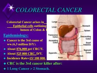
Colorectal cancer • Third most common type of cancer and second most frequent cause of cancer-related death • Estimated new cases in 2012 : 141,210 - 71,850 men and 69,360 women - 2/3 colon and 1/3 rectal cancers • Estimated deaths from Colorectal cancer in 2012 : 49,380 • Incidence rates: - High (Australia, New Zealand, Europe, US) - Low (are Africa and South-Central Asia) • A gradual shift toward right-sided or proximal colon cancers • Death rates from CRC have declined progressively since the mid-1980s
Colorectal Liver Metastases • The liver is the most common site of metastases in CRC patients . • 25% of CRC patients present with CLM (worse prognosis) • 30%-60% patients develop CLM. • 20%-30% patients develop Lung metastases
Colorectal Cancer (CRC) Sporadic (average risk) (75-80%) Family history(10-15%) Rare syndromes (<0.1%) Hereditary non-polyposis colorectal cancer (HNPCC) (3-5%) Familial adenomatouspolyposis (FAP) (1-2%)
Colorectal Cancer • 80% present with early disease • 20% present with metastatic disease. • Among patients diagnosed with early-stage disease, 40% will suffer recurrence Stage at Diagnosis Localized (Stage I/II) 40-45% Distant (Stage IV) 20-25% Regional (Stage III) 35%
5-Year Survival for CRC by Stage 70-90% 100 80 25-65% 65% 60 % of patients 40 20 5-16% 0 All Stages Localized Stage I ; II Regional Stage III Distant Stage IV
Risk Factors for CRC • Age >50 (average risk) • Racial, ethnic factors - African-Americans have increased risk • Dietary factors - High animal fat, low fiber diet • Lifestyle - Sedentary - Obesity - Smoking - Alcohol
Risk Factors for CRC • Family or personal history of CRC • HNPCC – Lynch syndrome I, II • Polyposis syndromes – FAP, Gardner’s syndrome, juvenile polyposis • Inflammatory bowel disease – chronic ulcerative colitis, Crohn’s disease
Protective factors • coffee consumption reduced risk of CRC • Physical activity • Diet • Fiber • Folic acid and folate • Calcium and Magnesium, vitaminD • Fish consumption • Drags: Aspirin and NSAIDs, Postmenopausal hormone therapy, Statins, Antioxidants
The Adenoma-Carcinoma process Mutations leading to formation of colorectal tumor
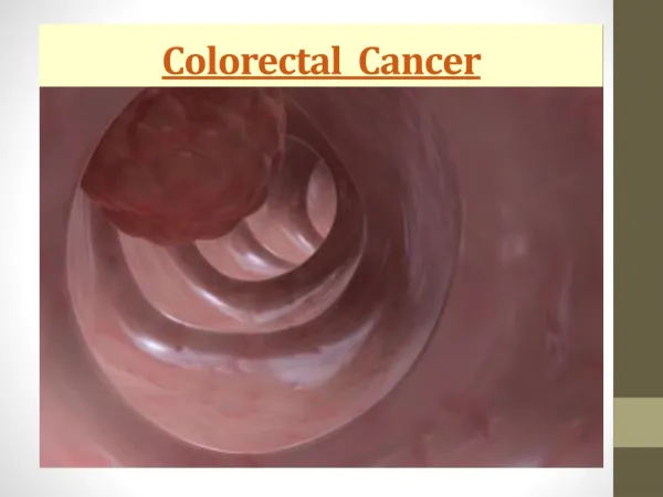
Symptoms ofColorectal Cancer • A change in bowel habits: diarrhea, constipation, or a feeling that the bowel does not empty completely • Bright red or dark blood in the stool • Stools that appear narrower or thinner than usual • Discomfort in the abdomen, including frequent gas pains, bloating, fullness, and cramps • Unexplained weight loss, constant tiredness, or unexplained anemia (iron deficiency) • Abdominal mass (late stage)
Unusual presentations • Local invasion causing malignant fistula formation into adjacent organs • Fever of unknown origin • Intraabdominal, retroperitoneal, or abdominal wall abscesses. • Streptococcus bovis bacteremia and Clostridium sepsis are due to underlying colonic malignancies in about 10 % of pts • CRC - 6 percent of adenocarcinomas of unknown primary sites
Staging Primary tumor • TX - Primary tumor cannot be assessed • T0No - evidence of primary tumor • Tis - Carcinoma in situ: intraepithelial or invasion of lamina propria • T1 - Tumor invades submucosa • T2 - Tumor invades muscularis propria • T3 - Tumor invades through the muscularis propria into pericolorectal tissues • T4a - Tumor penetrates to the surface of the visceral peritoneum • T4b - Tumor directly invades or is adherent to other organs or structures• Regional lymph node • NX - Regional lymph nodes cannot be assessed • N0No - regional lymph node metastasis • N1 - Metastasis in 1-3 regional lymph nodes N1a - Metastasis in one regional lymph node N1b - Metastasis in 2-3 regional lymph nodes N1c - Tumor deposit(s) in the subserosa, mesentery, or nonperitonealized pericolic or perirectal tissues without regional nodal metastasis • N2 - Metastasis in four or more regional lymph nodes N2a - Metastasis in 4-6 regional lymph nodes N2b - Metastasis in seven or more regional lymph nodes Distant metastasis • M0No - distant metastasis • M1 - Distant metastasis M1a - Metastasis confined to one organ or site (eg, liver, lung, ovary, nonregional node M1b - Metastases in more than one organ/site or the peritoneum
Pretreatment locoregional staging evaluation for colon cancer • Physical examination, rigid rectosigmoidoscopy • Carcinoembryonic antigen (CEA) • Full colonoscopy • CT colonography (virtual colonoscopy) • Double contrast barium enema • Computed tomography (CT) scans of abdomen and pelvis • positron emission tomography [PET] scan
Investigations • Double contrast barium enema • Does not require sedation • Avoids risk of perforation • More limited in detecting small lesions • Second line in patients who failed / cannot undergo colonoscopy
Investigations • Colonoscopy • Can visualize lesions < 5mm • Small polyps can be removed or at a later stage by endoscopic mucosal resection • Performed under sedation • Consent: bleeding, infection, perforation (1 in 3000), missed diagnosis, failed procedure, anaesthetic/medical risks • Warn: bowel prep, abdominal bloating/discomfort afterwards, no driving for 24 hours
CT colonography Advantage - Short procedure – 15- 20 minutes - No sedation or anesthesia - Non invasive - no risk of perforation of colon - Addition information about surround organ Disadvantage - Radiation - Expensive - Missing of small (<1cm) polyps - Unable to take biopsy Indication - Failed Colonoscopy - Elderly Frail Patients - Bleeding disorders - Obstructing Cancer - Sedation Issues - Screening
Polyps • 20% of population over 50 has adenomatous polyp • Patients with polyps are 5 times more likely to develop carcinoma • Patients with multiple polyps have a 2.5 times greater incidence of cancer that those with a single polyp • 2.5/1000polyps progress to carcinoma per year • Progression from adenoma to carcinoma takes approximately 5-10 years
Management of malignant polyp Benign adenomas, as well as those with severe dysplasia or carcinoma in situ (no evidence of invasive cancer) can be effectively managed by endoscopic removal (polypectomy) The presence of any of the following factors should prompt consideration of radical surgery: - Poorly differentiated histology - Lymphovascular invasion - Cancer at the resection or stalk margin - Invasion into the muscularis propria of the bowel wall (T2 lesion) - Invasive carcinoma arising in a sessile (flat) polyp with unfavorable features
Treatment • Surgery- primary • Chemotherapy • Radiotherapy • Targeted/immunotherapy
SURGERY In colorectal cancer the aims of surgery are: a. Excision of the tumor with adequate margin –5 cm of normal bowel proximal and distal to the tumor b. Removal of the lymph nodes that surround the colon and rectum and maycontain cancer cells c. Restoration of the continuity of colon d. Inspected other viscera for MTS
Management Cecum or ascending colon • Right hemicolectomy (RH) • Vessels divided – ICA and rt CA • Anastamosis between terminal ileum and transverse colon Transverse colon • Close to hepatic flexure RH • Mid-transverse extended RH • Splenic flexure SC or LH Descending colon • Left hemicolectomy (LH) • Vessels divided – IMA,LCA,SA
Management • Sigmoid colon • High anterior resection • Vessels ligated – IMS,LC,SA • Anastomoses of mid-descending colon to upper rectum • Obstructing colon carcinoma Right and transverse colon – resection and primary anastomosis Left sided obstruction • Hartmann’s procedure • Primary anastamosis – subtotal colectomy (ileosigmoid or ileorectalanastomosis) • Proximal diverting stoma • Palliative stent
First described 1991 Laparoscopic colorectal surgery • No difference in death rate • No significant difference in complications • At least equivalent oncologically to open colectomy Disadvantage - Longer time operations - Long learning curve - Port site metastasis Potential benefits • Smaller wounds • Less pain • Early return of bowel function • Early discharge • Early return to normal activities
Self Expanding Stents • Treatment of malignant large bowel obstruction - Inserted by colonoscopy under endoscopic and fluoroscopic control (or both) - Avoid emergency laparotomy with colostomy • Best for left side cancer - Right and transverse colon cancer can have resection and anastomosis - Rectal cancer rarely obstruct ( and stent migrated out) • Two situation - Palliative - “Bridge” to definitive surgery • Advantage - Safe and effective - Low mortality and morbidity - Lower costs
Drugsfor Treatment of Colorectal Cancer Chemotherapy • 5 Fluorouracil (5-FU) + Leucovorin – bolus or infusional • Capecitabine (Xeloda) • Irinotecan (CPT-11) • Oxaliplatin Biological therapy • Bevacizumab (Avastin) • Cetuximab (Erbitux) • Panitumumab
Adjuvant therapy • BASIS • Despite curative surgery half of these patients suffer INCURABLE TUMOR RECURRENCE leading to cancer related death • Therefore these is a need of adjuvant therapy to improve DFS and OS • Establishment of adjuvant therapy as a standard treatment in stage III colon cancer based on improvement in OS • In stage II colon cancer of adjuvant therapy remains conversial
Adjuvant therapy for colon cancer • STAGE I : Surgery only • Stage III: - FOLFOX- (5-FU+Leucovorin + oxaliplatin) - improved both OS and DFS • Adjuvant XRT: - for selected T4 lesion with penetration • Targeted therapy: - for advanced and metastatic disease • STAGE II : Adjuvant therapy controversial and considered for the following -obstructed or perforated colon ca - involvement of adjacent organ (T4 lesion) - high risk histology - inadequate LN sampling (<13) - elevated CEA - lymphovascular invasion - perineural invasion