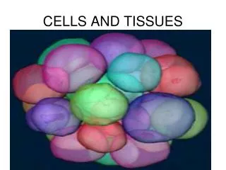
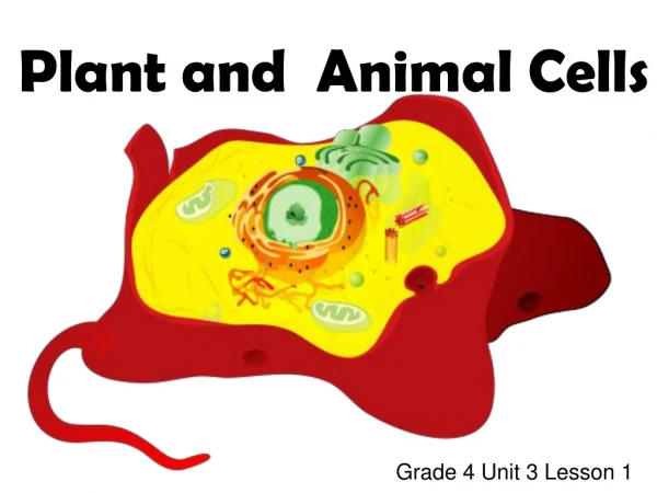
What Do SPECIALIZED Cells Do?
What Do SPECIALIZED Cells Do?. SEPUP Cell Biology: Activity 5. In the Activity : What D o Cells Do?, you examined some basic structures & organelles in cells . Many types of cells also have a specialized structure that allow the cells to perform specific functions. .

What Do SPECIALIZED Cells Do?
E N D
Presentation Transcript
What Do SPECIALIZED Cells Do? SEPUP Cell Biology: Activity 5
In the Activity : What Do Cells Do?, you examined some basic structures & organelles in cells. Many types of cells also have a specialized structure that allow the cells to perform specific functions.
For example, a muscle cell is specialized for movement, while a red blood cell is specialized for carrying oxygen throughout the body.
In this activity, you will examine the functions of some specialized cells. Challenge: What are the specialized structures & functions of cells
Use the instruction sheet to access the simulation for: What Do Specialized Cells Do? View each simulation for each specialized cell. As you view each simulation, label the organelles & structures on your mini-card. Answer the questions that go with each cell.
Cell #1: Neuron • Vesicles in the sending cell move to the membrane & release neurotransmitters into a space that has ions. The ions move to a place on the receiving cell’s membrane & enter the receiving cell. The flow of ions excites the receiving cell. • The cell is involved in sending a signal that excites another cell. • Chemical Synapse Simulation
Cell #2: Pancreatic • Insulin moves into a transport vesicle from the Golgi apparatus & fuses with another vesicle to make an even bigger vesicle. Next, glucose comes into the cell & the vesicle moves to the membrane where the insulin exits the cell. • The cell releases insulin to neighboring blood capillaries. • Diabetes Animation
Cell #3: Muscle • The myosin binds to the actin, causing the myosin to change shape & pull on the actin. The filaments slide past each other & the structure becomes shorter. There are lots of mitochondria near the filaments. • The muscle is contracting. • Each muscle cell is a single cell. The muscle cells make up muscle fibers: a bundle of smaller myofibrils made up of thin (protein actin) & thick (protein myosin) filaments. The thin filaments slide to make the muscle fiber contract. ATP is necessary to make this process occur!
Cell #4: Intestinal • The villi & microvilli are fingerlike projections from the cell membranethat increase the surface area for nutrients to absorb. Nutrients are moving from one side to another through intestinal absorptive cells that make up the villi & microvilli. • These intestinal cells move nutrients through the intestine into the capillaries of the blood.
Cell #5: Macrophage • The cell engulfs the microbe & contains in in a vesicle. The vesicle fuses with a lysosome & enzymes digest the microbe into small pieces. Some of these pieces are displayed on the cell membrane where they act as receptors for helper T cells. • The macrophage engulfs & digests microbes, such as bacteria, & helps other cells in the immune system to carry out their functions. • Macrophage Animation
Cell #6: Sperm • A set of microtubule doublets use ATP to crawl & cause bending of the bundle, which makes the tail whip back & forth. There is a group of mitochondria surrounding the segment of the microtubule doublets. • This cell is a sperm cell that can swim to an egg to fertilize! • Microtubules are straight, hollow tubes composed of globular proteins called tubulins. Microtubules are fibers that make up the cytoskeleton.

![[virtual] cells](https://cdn1.slideserve.com/3553683/slide1-dt.jpg)