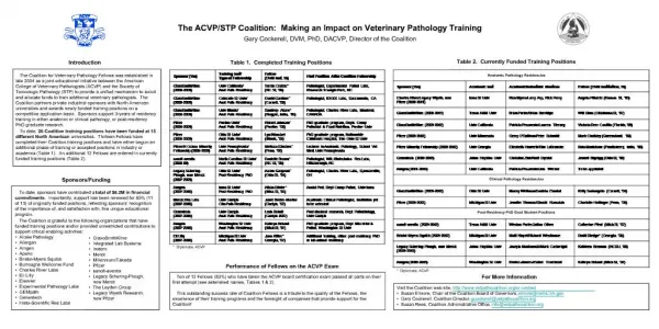
Download
1 / 40
420 likes | 1.29k Vues
Morphologic Classification of Mouse Mammary Tumors with Some Examples in Rat Sabine Rehm, Dr. med. vet., ACVP Diplomate SmithKline Beecham Pharmaceuticals. Outline. Classification of Neoplasms Classification of Rat Mammary Tumors Classification of Mouse Mammary Tumors 1950-90

E N D
Morphologic Classification of Mouse Mammary Tumors with Some Examples in RatSabine Rehm, Dr. med. vet., ACVP DiplomateSmithKline Beecham Pharmaceuticals
Outline • Classification of Neoplasms • Classification of Rat Mammary Tumors • Classification of Mouse Mammary Tumors 1950-90 • Histogenetic Classification of Mouse Mammary Hyperplasia and Neoplasia: Alveolar cell Ductal cell Myoepithelial cell
Overview: In the next 30 minutes, I will talk a little bit about classifications in general, then present what has been published recently for rat mammary tumors and briefly address terminology used for mice in the past 40 years or so. The main part of my talk will deal with a classification system based on cellular composition of mouse mammary tumors with regard to alveolar, ductal or myoepithelial differentiation.
Why Classify Neoplams? Clinician: prevention, detection, treatment, prognosis marker correlation, patient interest Academic Investigator: tumor biology defined by investigator interests FDA Regulator: hazard identification mechanism of tumor formation
The type of classification will depend on the goals to be achieved: The clinician is searching for markers that will correlate with treatment and prognosis, the main objective is the well-being of the patient. The academic investigator will most likely be interested in tumor biology and the focus will be on the interest of the investigator. Then there is the FDA regulator and this is where I am coming from. It is important to correctly classify lesion to allow hazard identification and mechanisms of tumor formation.
Morphologic Tumor Classifications Cellular Origin/Differentiation Cell type Grades of Malignancy Benign vs Malignant Subcategories Morphologic Variations
Morphologic Classification: Ideally, tumor classifications should be simple and easy to apply. However, much will depend on the experience of the person looking at a slide, since tumors inherently will assume a confusing array of morphologies. Basically tumors can be grouped according to cell type, such as epithelial vs mesenchymal, and according to malignancy. This would include simple hyperplasia, atypical hyperplasia, adenoma, carcinoma in situ and various types of carcinomas. Subcategories can be created according to distribution, subgrades of malignancy and morphologic variations . The extent of such subdivisions may depend on the goal of the classification system or personality of the investigator, being either a lumper or a splitter
Rat Mammary Tumors Russo et al. 1989van Zwieten et al. 1994 Epithelial Cystadenoma/Adenoma Adenoma Papilloma Carcinoma Adenocarcinoma Epithelial-stromal Fibroadenoma Fibroadenoma Adenolipoma Carcinosarcoma Carcinosarcoma Stromal Fibroma Fibroma Fibrosarcoma
Rat Russo vs van Zwieten: Comparing two recently published classifications of the rat mammary gland by Russo et al and van Zwieten et al, there is agreement on the main categories of epithelial, epithelial-stromal and stromal tumors but there are several differences. There are benign adenomas in both classifications but papillomas only in one. Malignant epithelial tumors on the one side are referred to as carcinomas and on the other side are called adenocarcinomas. This difference in nomenclature is probably one we will also have to deal with in this workshop. To me, carcinoma is suggestive of ductal differentiation, whereas adenocarcinoma implies glandular alveolar cell differentiation. Fibroadenomas constitute the vast majority of spontaneous mammary tumors in rats. In rats treated with chemical carcinogens, malignant tumors probably prevale and we will hear more on this subject from Dr. Russo. Carcinosarcomas in rats are mostly derived from fibroadenomas, as evidenced by neoplasms clearly showing the transition from benign to malignant. Neoplastic spindle cells can also be formed as an extreme dedifferentiation from epithelial cells or represent myoepithelial cells, as I will show you in a while.
Classification Rat vs Mouse Mammary Tumors Epithelial Rat Mouse Benign Malignant Epithelial-myoepithelial (B/M) Epithelial-stromal Benign (fibroadenoma) ? Malignant (carcinosarcoma) ? Stromal Benign (fibroma) Malignant (fibrosarcoma)
Rat vs Mouse: Now let us compare mammary tumors of rats and mice. In both pure epithelial malignant neoplasms outweigh benign adenomas, based on local invasion and cytologic appearance. Metastases, will depend on the model. Rodents are usually killed because of tumor size and skin erosions with associated bleeding. In rats, there is no special category for tumors with myoepithelial cells, although these tumors have been reported to occur in rats. Old mice spontaneously develop benign adenomyoepitheliomas, and malignant myoepitheliomas can reliably be induced by DMBA. As mentioned, epithelial-stromal or pure stromal tumors are common in rats, but rare in mice. However, in the transgenic slide set, there are probably some than can be called fibroadenomas. Many carcinosarcomas diagnosed to date in mice, probably represent malignant adenomyoepitheliomas.
Mouse Mammary Tumor Classification 1950-1990 Morphological and Alphabetical Adenocacrcinomas Type A Type B Type A/B Type C Type Y Type L Pale cell Fissured Adenoacanthoma, Organoid/Molluscoid Tumor, Carcinoma type P, Carcinosarcoma, Fibroadenoma, Mixed Tumor
1950-1990: Previous classifications of mouse mammary tumors represented a combination of morphologic structures and the alphabet. It is not a systematic evaluation, it is difficult to understand, there are many oldfashioned names such as adenoacanthoma or moluscoid tumor and there is considerable overlap. This is not a useful approach.
Determining Cellular Differentiation Stem - Alveolar - Ductal - Myoepithelial - Fibroblastic - Multiple Cell Types • Fixation: formalin 10% neutral buffered • Processing artifacts • Initial emphasis: small, early lesions • Cellular markers/Additional techniques • DDX: salivary/lacrimal glands, dermal elements, clitoral gland, lung tumors
Cellular differentiation: How do we obtain a useful system? At first we can try to go on the path of determining the cellular differentiation of a tumor. There are clearly limits to this, but at least we can give it a try. In the transgenic slide set, there were good examples of early and late lesions, but there are also slides with problems. Initially, the best fixation for H&E slides is formalin and beware of processors that do not perform properly. The tissue is permanently damaged. It is very helpful to first concentrate on early lesions that are not yet visible macroscopicallyLocalization of initial lesions will often enable us to determine the origin of larger neoplasms. Initially, cells may still retain many differentiated features or markers that will help characterize the cell types. Normal mammary tissue can be expected to be present in the same section, serving as an internal positive control for markers. In larger tumors, the origin is obscured due to invasive growth, cells have dedifferentiated losing markers or tissue is lost altogether because of widespread necrosis. One also has to keep in mind, that not all sc nodules arise from the mammary gland. In the slide collection there appears to be a tumor of the parotid gland – or tumors can arise from the skin. When it comes to assessing pulmonary metastases, one needs remember that mice have a reputation for developing lung tumors and we a couple of those in the slide set too.
Basal lamina Ductal cell Alveolar cell Myoepithelial cell MILK DUCT ACINUS Lobule of active gland Normal Mouse Mammary Gland
Mammary Gland cartoon: Tumors can arise from one or more cell types retaining features typical or even specific for the cell of origin. The mammary gland of the mature mouse is composed of ducts and acini embedded in a stroma consisting of fat cells, connective tissue and other supportive structures. The acinus is lined by alveolar cells, also referred to as acinar cells, producing milk fat and proteins when stimulated and the ducts are lined by ductal epithelium Both units are surrounded by myoepithelium enclosed by a basal lamina. Myoepithelial cells are contractile cells responsible for advancing milk produced by the system. Well formed lobules composed of numerous acini are only present in the mouse during lactation. I do understand that the term 'lobular' has by tradition a specific meaning in the context of the human breast. However, for the time being, I will avoid using the term lobule or lobular since it implies to me an unspecific gross or subgross structural organization which is also used in other organs, eg, in the liver. Lobules are always composed of several different cell types and therefore it is a rather unspecific term. This is probably something we can discuss later on.
Alveolar cell Myoepithelial cell Ductal cell Mouse Mammary TumorsRehm & Liebelt 1996 • Alveolar differentiation: Adenoma Adenocarcinoma
Rehm & Liebelt classification: I will take you now through a classification system of mouse mammary tumors based on cellular differentiation. Firstly, there are lesions with alveolar cell features, which I assume are the equivalent to human lobular lesions. These represent the majority of mammary tumors in mice, with or without mammary tumors virus, and were also commonly represented in the slide set.
Alveolar Hyperplasia • Focal /Multifocal/ Diffuse • Atypical • Increased serum prolactin levels • Cytoplasmic lipid droplets & secretions • Squamous metaplasia
TUMOR GROWTH PATTERNS TUBULAR CRIBRIFORM ACINAR SOLID PAPILLARY COMEDO NESTS TRABECULAE SHEETS
Tumor growth patterns: This cartoon summarizes different growth patterns mouse mammary adenocarcinomas can assume. Variations range from the well-differentiated acinar pattern to tubular, papillary, comedo and solid, forming sheets, trabeculae or nests. A single tumor can display one or even all of these features. Acinar structures can be absent and then it becomes difficult to classify the tumor. In such instances it may be useful to have an additional category, such as carcinoma NOS, meaning not otherwise specified.
Alveolar cell Myoepithelial cell Ductal cell Mouse Mammary TumorsRehm & Liebelt 1996 Alveolar differentiation: Adenoma Adenocarcinoma Adenosquamous carcinoma
Alveolar cell Myoepithelial cell Ductal cell Mouse Mammary TumorsRehm & Liebelt 1996 Alveolar differentiation: Adenoma Adenocarcinoma Adenosquamous carcinoma Ductal differentiation: Ductal papilloma/carcinoma
Ductal Hyperplasia • Intraductal • Ductal (budding/branching) • Focal/Multifocal/ Diffuse • Atypical
DUCTAL HYPERPL: • Obvious intraductal hyperplasia is easily recognized, whereas, ductal hyperplasia that occurs through budding and branching, may not and certainly needs to be distinguished from alveolar hyperplasia.
DUCTAL CARCINOMA: • It seems logical to call the neoplasm that arises from an intraductal hyperplasia, an intraductal carcinoma. However, since ductal tumors could also form by branching and budding, the term intraductal may be too limiting. It may be easier to call these lesions simply ductal carcinomas.
Alveolar cell Myoepithelial cell Ductal cell Mouse Mammary TumorsRehm & Liebelt 1996 Alveolar differentiation: Adenoma Adenocarcinoma Adenosquamous carcinoma Ductal differentiation: Ductal papilloma/carcinoma Ductal sqaumous carcinoma
DUCTAL SQUAMOUS CARCINOMA: • Similar to the adenosquamous carcinoma, there is a ductal squamous carcinoma. • Ductal squamous carcinomas can be induced with DMBA. They are rather peculiar tumors since they have this radiating growth pattern. At first sight, one may think of a skin tumor which is not unusual since the mammary gland is an adenexal gland of the skin. Therefore, it is not surprising to be reminded of this fact in neoplasms. These lesions, however, have no contact with the skin.
Alveolar cell Myoepithelial cell Ductal cell Mouse Mammary TumorsRehm & Liebelt 1996 Alveolar differentiation: Adenoma Adenocarcinoma Adenosquamous carcinoma Ductal differentiation: Ductal papilloma/carcinoma Ductal sqaumous carcinoma Myoepithelial differentiation: Adenomyoepithelioma (Benign/Malignant)
ADENOMYOEPITHELIOMA: The third category of mouse mammary tumors shows myoepithelial differentiation. Similar tumors are known to occur in humans and I suspect there are several in the transgenic slide set. This is my special field of interest where I was able to do some experimental work at NCI.
Mouse DMBA-induced Mammary Tumors Study Design: Strains: B6D2F1 or D2B6F1 (C57BL x DBA) Age at start: 8 weeks Compound: 7,12-dimethylbenz()anthrazene Dose: 1 mg/mouse Dosing: 1/week for 4 weeks
Mouse DMBA-induced Mammary Tumors Results: 6-8 weeks after last treatment: ductal squamous carcinomas 6-8 months after last treatment: adenomyoepitheliomas
Transplantation of Adenomyoepitheliomas and Clonally Selected Malignant Myoepithelium Hypothesis: 1. Cellular differentiation potential of myoepithelial cells (cartilage/bone) 2. Increase capability to metastasize
Classification of Mouse Mammary Tumors • Epithelial, benign: Adenoma/ Papilloma/Fibroadenoma • Epithelial, malignant: Adenocarcinoma Adenosquamous carcinoma Ductal carcinoma Ductal sqaumous carcinoma • Myoepithelial tumors: Adenomyoepithelioma (Benign/Malignant) • Carcinoma, NOS
Final Classifcation: In summary, it can be concluded that most proliferative lesions of the mouse mammary gland can be classified into benign epithelial, some epithelial-stromal, and malignant epithelial processes. These lesions can show alveolar or ductal differentiation with or without a dominant squamous component. Very rarely neoplasms may also show both alveolar and ductal differentiation. Thirdly, there is a less common category composed of myoepithelium and various other cellular elements. And if you have no idea on the differentiation of the neoplasm, there is always carcinoma,NOS