Download
1 / 39
420 likes | 841 Vues
Chapter 17a Circulation Characteristics of Blood RBC and Erythropoiesis. Overview of Vessels. Veins: vessels that move TOWARD the heart. Arteries: blood vessels that move AWAY from the heart. Capillaries: Small vessels created by repeated branching of arteries and veins

E N D
Chapter 17aCirculationCharacteristics of BloodRBC and Erythropoiesis
Overview of Vessels Veins: vessels that move TOWARD the heart Arteries: blood vessels that move AWAY from the heart • Capillaries: Small vessels created by repeated branching of arteries and veins • 1 epithelial cell layer thick • allows for gas, waste, and nutrient exchange at the tissue level
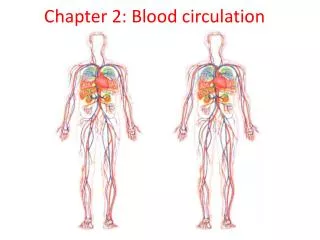
Blood Circulation 5. O2 and nutrients diffuse across capillary walls into tissues 6. CO2 and waste move from tissues into vessels 4. Blood leaves left ventricle of the heart via aorta and moves into arteries towards tissues 1. O2 deficient blood moves into veins that enter the right atrium of heart 2. Right ventricle pumps blood through the pulmonary artery, which carries blood to lungs, where it releases CO2 and picks up O2 3. O2 rich blood returns to the heart via pulmonary veins and enters left atrium of heart
Heart Anatomy (pump your blood song) Aortic Valve = L Semi-lunar Valve Pulmonary Valve = R Semi-lunar Valve LAtrioventricular valve = Mitral Valve = Bicuspid Valve R Atrioventricular valve = Tricuspid valve
Physical Characteristics of Blood • Body’s only fluid tissue • Sticky plasma • Color: scarlet (O2 rich) to dark red (O2 poor) • pH: 7.35–7.45 (slightly basic) • Temperature: 38C (100.4F) • 8% of total body weight • AVG volume: • males: 5 - 6 L • females: 4 - 5 L
Functions of Blood • Distribution: Transportation of • Oxygenfrom lungs to cells • Nutrientsfrom intestines to cells • Metabolic wastesfrom cells to lungs and kidneys for elimination • Hormones from endocrine glands to target organs
Functions of Blood 2. Regulation: Maintenance of • Body temperature • pH with buffer proteins and solutes • pH imbalances interfere w/ cell activities, causing tissue damage • Fluid volume
Functions of Blood 3. Protection: • Fights infections • Synthesizing and employing antibodies • Activating complement proteins • Activating WBCs to destroy foreign invaders
Functions of Blood 3. Protection: b. Prevention of blood loss by • Activating plasma proteins and platelets • Initiating clot formation when a vessel is broken
Composition of Blood • Composed of liquid plasma (matrix) and formed elements (cellular)
Composition of Blood • Hematocrit = % of RBCs out of total blood volume Males: 47% Females: 45%
Blood Plasma • 90% water by volume • > 100 solutes, including: 1. Proteins • Albumin (liver protein): • blood buffer; maintains osmotic pressure • Globulins: • α, β (liver proteins) : transport lipids, metal ions, fat-soluble vitamins • Gamma ɣ: antibodies during immune response • Fibrinogen: clotting protein
Blood Plasma • 90% water by volume • > 100 solutes, including: 2. Lactic acid, urea, creatine 3. Hormones: ex. insulin, erythropoietin, and others 4. Organic nutrients: carbohydrates, amino acids, lipids 5. Electrolytes: Na+, K+, CA2+,Cl-, bicarbonate (HCO3), etc. 6. Respiratory gases: O2 and CO2
Formed Elements • Erythrocytes (RBC), leukocytes (WBC), and platelets (for clotting) • Only WBCs are complete cells • RBCs have no nuclei or organelles • platelets are just cell fragments • Most survive in the bloodstream for only a few days (RBC: 100 – 120 days) • Most are amitotic (do not divide) • replaced by stem cells in bone marrow
Erythrocyte (RBC) Characteristics • Biconcave discs High SA/V ratio (> 30% than spherical) • Contributes to blood’s viscosity • anucleate (no nucleus), and no organelles, no mitochondria • anaerobic ATP generation; do not use any O2 that they transport • Contain spectrin membrane protein on cytoplasmic face • Flexibility • Ability to change shape • Ease of movement through narrow capillaries
Gas Transport • RBCs with > 97% hemoglobin (Hb) • protein used for transport of O2 and CO2 • Binding Hb to CO arrests cellular respiration
Hemoglobin (Hb) Structure • 4 Globin proteins, each globin bound to a hemegroup • Heme = red pigment w/ an atom of iron (Fe) • binds to O2 and changes to scarlet red • Fun Fact: • 1 Hb can transport 4 O2molecules • One RBC has 250 million Hbmolecules • The cell transports 1 billion molecules of O2!
Forms of Hb • Oxyhemoglobin – Hb bound to oxygen O2 • O2loading happens in the lungs • Deoxyhemoglobin – Hb after O2 diffuses into tissues • Carbaminohemoglobin – Hb bound to CO2 • CO2 loading happens in the tissues How does loading and unloading of respiratory gasses happen? Watch this video and take notes to find out…
Hematopoiesis • Blood Cell Formation • Occurs in red bone marrow of: • Axial skeleton and girdles • Epiphyses of the humerus and femur • Hemocytoblasts (adult stem cells of blood): • - give rise to all formed elements • Erythropoeisis: specific formation of RBC
Erythropoiesis • Developmental Pathway • Phase 1: Ribosome synthesis for globulin protein formation • Phase 2: Hemoglobin accumulates • Phase 3: Nucleus and Organelles eject • Reticulocytes: still w/ remnants of RER and ribosomes • Erythrocytes: complete RBC, all ribosomes degraded by enzymes • Fun Fact: process takes 15 days; 2 million RBCs made/minute
Dietary Needs 1. Iron (Fe) • 65% of Fe in body is in Hb • Remainder stored in liver, spleen, marrow as protein-iron complexes: ferritin and hemosiderin • free iron ions Fe2+ and Fe3+ are toxic, increasing free radicals, causing cell death 2. vitamin B12and folic acid • For normal DNA synthesis 3. Lipids, CHO, Amino Acids Iron lost daily via waste • 0.9 mg/day lost by men • 1.7 mg by women (menstrual bleeding)
Control of Erythropoiesis • Delicate balance btw RBC formation and destruction • Too few RBC hypoxia (lack of O2 in tissues) • Too many RBC blood is too viscous • Hormonal Controls • Erythropoietin (EPO): • hormone made by kidneys • directly stimulates RBC formation • EPO released in response to: • Tissue hypoxia • Increased tissue demand for O2
EPO Mechanism Imbalance Start Stimulus: Hypoxia due to decreased RBC count, decreased amount of hemoglobin, or decreased availability of O2 Homeostasis: Normal blood oxygen levels Imbalance Increases O2-carrying ability of blood Reduces O2 levels in blood Kidney (and liver to a smaller extent) releases erythropoietin Erythropoietin stimulates red bone marrow Enhanced erythropoiesis increases RBC count
Fate of Erythrocytes • RBC w/ no nuclei, no DNA, no new proteins • Fragile, lose shape, becomes rigid • Hb degenerate • RBCs often trapped and fragmented in spleen • Dying RBC engulfed and destroyed by macrophages • Components of Hb are broken down and some parts recycled for reuse.
Life Cycle of Erythrocytes Low O2 levels in blood stimulate kidneys to produce erythropoietin. Erythropoietin levels rise in blood. Erythropoietin and necessary raw materials in blood promote erythropoiesis in red bone marrow. New erythrocytes enter bloodstream; function about 120 days. Aged and damaged red blood cells are engulfed by macrophages of liver, spleen, and bone marrow; the hemoglobin is broken down. Hemoglobin
Destruction of Hb 1. Heme and Globin separated Hemoglobin Heme Globin
Destruction of Hb 1. Heme and Globin separated Hemoglobin Heme Globin 2. Globin metabolized into AA and released into blood Amino acids
Destruction of Hb 1. Heme and Globin separated Hemoglobin Heme Globin 2. Globin metabolized into AA and released into blood 3. Heme degraded into yellow pigment Bilirubin Bilirubin Amino acids Iron stored as ferritin, hemosiderin
Destruction of Hb 1. Heme and Globin separated Hemoglobin Heme Globin 2. Globin metabolized into AA and released into blood 3. Hemedegaded into yellow pigment Bilirubin Bilirubin Amino acids Iron stored as ferritin, hemosiderin 4. Iron stored safely as ferritin and hemosiderin in liver
Destruction of Hb 1. Heme and Globin separated Hemoglobin Heme Globin 2. Globin metabolized into AA and released into blood 3. Hemedegaded into yellow pigment Bilirubin Bilirubin Amino acids Iron stored as ferritin, hemosiderin 4. Iron stored safely as ferritin and hemosiderin in liver 5. Iron is bound to transferrinprotein and released into circulation for erythropoiesis
Destruction of Hb 1. Heme and Globin separated Hemoglobin Heme Globin 2. Globin metabolized into AA and released into blood 3. Hemedegaded into yellow pigment Bilirubin Bilirubin Amino acids Iron stored as ferritin, hemosiderin 4. Iron stored safely as ferritin and hemosiderin in liver 5. Iron is bound to transferrin protein and released into circulation for erythropoiesis 6. The liver secretes bilirubin into intestine with bile from the gall bladder 7. The intestines metabolize it, and it leaves the body in feces as a pigment called stercobilin (brown poop) Bilirubin is metabolized by intestines and excreted in feces
Destruction of Hb 1. Heme and Globin separated Hemoglobin Heme Globin 2. Globin metabolized into AA and released into blood 3. Hemedegaded into yellow pigment Bilirubin Bilirubin Amino acids Iron stored as ferritin, hemosiderin 4. Iron stored safely as ferritin and hemosiderin in liver 5. Iron is bound to transferrin protein and released into circulation for erythropoiesis 6. The liver secretes bilirubin into intestine with bile from the gall bladder 7. The intestines metabolize it, and it leaves the body in feces as a pigment called stercobilin (brown poop) Bilirubin is metabolized by intestines and excreted in feces Circulation Food nutrients, including amino acids, Fe, B12, and folic acid are absorbed from intestine and enter blood 8. Raw materials are made available in blood for erythrocyte synthesis.
Erythrocyte Disorders • Polycythemia – excess RBCs that increase blood viscosity • Causes clotting in small capillaries, leading to • Stroke • Heart Attack • 3 types: • Polycythemia vera: bone marrow disease • 20 polycythemia: people living at high altitudes. • Blood doping : enhancing RBC numbers to increase athletic performance • removing blood for a few days, then re-infusing it.
Erythrocyte Disorders 2. Anemia– blood has abnormally low oxygen-carrying capacity • Symptom; not a disease • Blood oxygen levels cannot support normal metabolism • Causes fatigue, paleness, shortness of breath, chills
Anemia: Insufficient Erythrocytes • Hemorrhagic anemia: due to acute or chronic loss of blood (ex. stab wounds) • Hemolytic anemia: RBCs prematurely destroyed (ex. abnormal proteins of RBCs, abnormal immune system, parasitic) • Aplastic anemia: bone marrow failure: does not produce enough RBCs
Anemia: Decreased Hb Content • Iron-deficiency anemia results from: • hemorrhagic anemia • Poor diet: lack of iron-containing foods • Impaired iron absorption • Pernicious anemia results from: • Inability to absorb vitamin B12 • Not enough intrinsic factor (protein that aids in B12 absorption)
Anemia: Abnormal Hemoglobin • Thalassemias – absent or faulty globin chain in Hb • RBCs are thin, delicate, and deficient in Hb • Can lead to hemolytic anemia • Sickle-cell anemia – from abnormal Hb-S • Hb-S due to genetic mutation of a single amino acid (substitution), causing sickle-shape of RBC • Both conditions offered some protection from malaria