Download

1 / 42
450 likes | 725 Vues
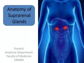
SUPRARENAL GLANDS. Suprarenal or adrenal glands are a pair of small golden yellowish bodies situated retroperitoneally anterosuperior to superior renal pole. COMPARATIVE ANATOMY. In anurans adrenals are flattened elongated bodieslocated against ventral surfaces of kidney.

E N D
Suprarenal or adrenal glands are a pair of small golden yellowish bodies situated retroperitoneally anterosuperior to superior renal pole.
COMPARATIVE ANATOMY • In anurans adrenals are flattened elongated bodieslocated against ventral surfaces of kidney. • In urodeles they lie near medial border. • In amniotes they are near cephalic pole of kidney.
Mammalian adrenals consist medulla and cortex but in fishes they are not part of same organ.
Medulla • In fishes medullary cells form segmental glandular masses near autonomic ganglia. • In tetrapods they aggregate on or near kidney and are intimately associated with cortical component.
Cortex • In fishes a series of paired glandular masses arises from the coelomic mesoderm and assumes a position between kidneys and post cardinal veins as interrenal bodies. • In amphibians , reptiles and birds the interrenal tissue is interspersed among the ectodermal medullary masses forming a single mass of dual organ.
In mammals the interrenal tissue now called adrenal cortex completely surrounds the medullary component. • Accessory cortical tissue may remain apart from main gland in all tetrapods.
DEVELOPMENT • Developed by 2 sources – Cortex – mesodermal. Medulla – Ectodermal.
Cortex – develops at 37 days from columnar mesothelial cells lining the coelomic cavity in the angle between root of mesentry and developing gonad. • Medulla – composed of cells derived from the neural crest cells which reach the mediodorsal aspect of the gland at 44 days and begins to invade it.
Capsule is completely formed in the later half of fetal life. • Catecholamines appear by 10th week of fetal life. • Glomerular zone is poorly developed in fetus.
Fetal suprarenals are relatively larger. It is due to extensive development of cells in fetal cortex. • Probable causes – > Stimulated by ACTH from fetal pituitary. > Human chorionic gonadotropin from placenta.
Accessory suprarenal tissue – Cortical tissues found > around main gland. > within broad ligament of uterus. > within spermatic cord or testis. > within mesentries of gut.
GROSS ANATOMY. • Weight – 5g. At birth – 1/3rd of kidney. Adult – 1/30th of kidney. • Size – vertical – 50mm transverse – 30mm anteroposterior – 10mm.
Capsule 1] Inner true capsule. 2] Outer false capsule derived from renal fascia.
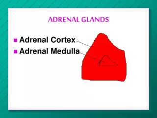
Right gland • Shape – Irregular tetrahedron or triangular or pyramidal. • Presenting parts – Base. Apex. Anterior surface. Posterior surface. Medial border. Lateral border.
Left gland. • Shape – Semilunar. • Presenting parts – Anterior surface. Posterior surface. Medial border. Lateral border. Upper end. Lower end.
Structures between medial border of 2 glands – • Right crus with right celiac ganglion and right inferior phrenic artery. • Azygous vein. • Cisterna chyli and thoracic duct. • Abdominal aorta with celiac artery. • Left crus with left celiac ganglion and left inferior phrenic artery.
STRUCTURE. • Gross section of a fresh gland shows – > outer broader yellowish zone of cortex. > Inner dark red / pearly grey colored medulla. Because of irregular shape the line of separation between cortex and medulla is irregular, distribution of medulla also varies.
CORTEX. • Divided into 3 zones • Outer subcapsular zone – zonaglomerulosa which is a relatively narrow zone showing small polyhedral cells in rounded groups or curved columns.
2 ] Middle zone – zona fasciculata is the broadest zone consisting of large polyhedral cells which course parellel to one another in a radial direction towards medulla. Cytoplasm contains abundant fat droplets composed of cholesterol, fatty acids.
3 ] Inner zone – zona reticularis made up of branching interconnected column of round cells. Cytoplasm has few lipid droplets.
Medulla – Composed of groups and columns of chromaffin cells. Cells are columnar in shape and the chromaffin reaction is due to their content of catecholamines. 2 types of cells – 1] Norepinephrine cells stain darkly with silver and iodine. 2] Epinephrine cells stain lightly / will not take stain.
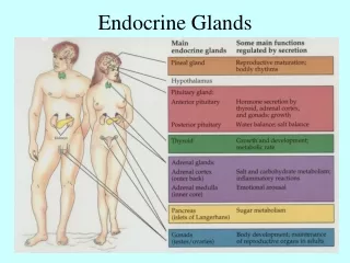
HORMONES SECRETED. • Zona glomerulosa:- Secretes mineralocorticoids- promote sodium retention and potassium excretion from kidneys. 1] Aldosterone. 2] Deoxy corticosterone – 3% effect of aldosterone.
Zona fasciculata and reticularis:- Secretes glucocorticoids and sex steroids. • Glucocorticoids- acts on carbohydrate and protein metabolism. Also has mineralocorticoid activity. 1] Cortisol. 2] Corticosterone – weak glucocorticoid.
Sex steroids- 1] Androgen – activity is less than 20% of testosterone. 2] Oestrogen and progesterone in very small amounts.
Secretions are mainly controlled by adrenocorticotrophic hormones released by hypophysis cerebri.
Baroreceptor discharge Emotions Trauma Hypothalamus/ pituitary Adrenal cortex. Cortisol
Medulla – Secretes adrenaline & noradrenaline.
BLOOD SUPPLY. Supplied by 3 arteries • Superior suprarenal artery branch of inferior phrenic artery. • Middle suprarenal artery branch of abdominal aorta. • Inferior suprarenal artery branch of renal artery.
Medullary vein emerges from the hilum to form a suprarenal vein which drains into inferior venacava on right side and left renal vein on left side. • Right suprarenal vein is much shorter.
Lymphatic drainage – To lateral aortic lymph nodes.
Nerve supply – Medulla is supplied by the sympathetic nerves from celiac plexus conveying fibres from T8 – L1 segments of spinal cord. Cortex is controlled by hypophyseal Adrenocorticotrophic hormone.
APPLIED ASPECT. • Atrophy of the cortex – Addison’s disease. • Adrenal hyperplasia – > Excess secretion of glucocorticoids – Cushing’s syndrome. > Excess secretion of mineralocorticoids – Conn’s syndrome.
Adrenogenital syndrome. • Female pseudohermaphroditism. • Tumour of medulla - Pheocromocytoma. • Adrenalectomy.