Download
1 / 35
500 likes | 1.7k Vues
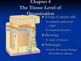
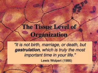
The Tissue Level of Organization. "It is not birth, marriage, or death, but gastrulation , which is truly the most important time in your life." Lewis Wolpert (1986). The Tissue Level of Organization. Group of similar cells common embryonic origin common function

E N D
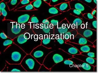
The Tissue Level of Organization "It is not birth, marriage, or death, but gastrulation, which is truly the most important time in your life." Lewis Wolpert (1986)
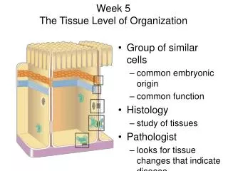
The Tissue Level of Organization • Group of similar cells • common embryonic origin • common function • bound together by intercellular substance • Histology • study of tissues What kind of molecules may make up these intercellular junctions?
The Origin of Tissues Differentiate between primary tissues and embryonic germ layers. Morula Blastula Gastrula
Gastrulation in birds and mammals from LIFE: The Science of Biology, Purves et al, 1998 Does this process result in a change in the shape of the embryo? Does it change the number of embyronic germ layers?
Define each of these terms… List at least on specialization from each embryonic germ layer.
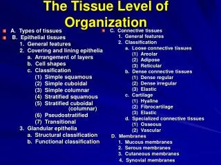
4 Basic Tissues Types • Epithelial • Connective • Muscle • Nervous
Intercellular Junctions • Tightjunctions • Adherens junctions • Gapjunctions • Desmosomes • Hemidesmosomes What does “hemi” mean?
Tight Junctions This type of intercellular junction is common in transport epithelia. • Why do you think tight junctions are located nearer the luminal cell border rather than the basolateral border? • Describe the structure and function of the apical cell border for these cells that line the intestine. • Where are transport epithelia found?
Gap Junctions What kinds of materials may pass between cells through gap junctions? In which tissues are gap junctions common?
Desmosomes • Give at least two examples of tissues containing desmosomes. • Where else would you expect to find keratin, and for what purpose? • What analogy would you use to describe the structure of a desmosome? • What function does this structural feature provide for the tissue?
Hemidesmosomes • Half of a desmosome! • Connect cells to extracellular material • basement membrane Which tissues are connected to a basement membrane, and would therefore possess hemidesmosomes?
Epithelial Tissue -- General Features • Cover surfaces, line cavities and form glands • Name one multicellular gland formed by epithelial tissue. • Differentiate between endocrine, exocrine and heterocrine glands. • Attached to underlying connective tissue by a basement membrane • Avascular---without blood vessels • nutrients diffuse in from blood vessels in underlying connective tissue • What does this mean for especially thick epithelia? • Good nerve supply • Rapid cell division; responsive to environmental stresses • Named according to the shape and arrangement of cells • List the general functions of epithelial tissues.
Epithelial Tissues and Their Basement Membrane What function might the basement membrane serve in the repair of injury to the epithelium?
Simple Squamous Epithelium Single layer of flat cells • lines blood vessels (endothelium), closed body cavities (mesothelium) • very thin --- controls diffusion, osmosis and filtration What is the primary function of this tissue (protection or transport)? (Hint: What is its function as an endothelium,…as mesothelium?)
Section of intestine showing serosa Surface view of lining of peritoneal cavity Examples of Simple Squamous Epithelium Is this endothelium, mesothelium, or neither?
Connective Tissues • Cells rarely touch due to usually large amount of intercellular material (extracellular matrix) • Matrix(fibers & ground substance) secreted by cells • Consistency varies from liquid or gel to solid • Function is to support, connect, protect and insulate • Good nerve & blood supply except cartilage & tendons Areolar c.t. What are the three major cell types often found in connective tissues, and what are their functions?
Collagen (25% of protein in your body) tough, resistant to pull, yet pliable Elastin (lungs, blood vessels, ear cartilage) Reticular (spleen and lymph nodes) thin, branched fibers that form framework within organs formed from protein collagen Types of Connective Tissue Fibers • smaller diameter fibers formed from protein elastin surrounded by glycoprotein (fibrillin) • can stretch up to 150% of relaxed length and return to original shape
Embryonic Connective Tissue:Mesenchyme • Irregularly shaped cells • In semi-fluid ground substance with reticular fibers • Gives rise to all other types of connective tissue
Areolar (loose) Connective Tissue • Black, fine = elastic fibers, • Pink, thick = collagen fibers • Purple Nuclei are mostly of fibroblasts What happens to the characteristics of this tissue if it becomes dehydrated?
Adipose Tissue • Peripheral nuclei due to large fat storage droplet • Deeper layer of skin, organ padding, yellow marrow • Reduces heat loss, energy storage, protection Why are these cells sometimes called “signet ring” cells?
Dense Regular Connective Tissue • Collagen fibers in parallel bundles with fibroblasts between bundles of collagen fibers • White, tough and pliable when unstained (forms tendons) • Also known as white fibrous connective tissue Do you see many blood vessels in this tissue? Implication?
Hyaline Cartilage • Chondrocytes sit in spaces called lacunae • No blood vessels or nerves so repair is very slow • Matrix may or may not contain fibers What would you call the cells that form this tissue?
Compact Bone • Osteon = lamellae (rings) of mineralized matrix • calcium & phosphate---give it its hardness (________________) • interwoven collagen fibers (and other proteins) provide strength and flexibility (______________) • Osteocytes in spaces (lacunae) in between lamellae • Canaliculi (tiny canals) connect cell to cell • Central canal contains blood vessels, nerves & a lymphatic vessel
Blood • Connective tissue with a liquid matrix = the plasma • Formed elements = red blood cells (erythrocytes), white blood cells (leukocytes) and cell fragments called platelets • Provide clotting, immune functions, carry nutrients, wastes, etc. What are the functions of each of the formed elements? Where are the formed elements manufactured?
Muscle • Cells that shorten due to the chemical and physical interaction between myofilaments • Actin and Myosin Compare and contrast this feature of muscle tissue cells with other cells not specialized for contraction. • Types of muscle • skeletal muscle • cardiac muscle • smooth muscle Compare the functions of the three types of muscle tissue. How is contraction in each of these tissues controlled?
Skeletal Muscle • Cells are large long cylinders with many peripheral nuclei How did these cells become multinucleate? • Visible light and dark banding (looks striated) What structure(s) within these cells account for the striations? • Voluntary (conscious) control
Cardiac Muscle • Cells are branched cylinders with one central nuclei, striated • Involuntary If so, then why are there nerves that innervate the heart? • Attached to and communicate with each other at intercalated discs What type(s) of intercellular junctions are present at intercalated discs?
Smooth Muscle • Spindle shaped cells with a single central nuclei • Walls of hollow organs (blood vessels, GI tract, bladder), often arranged in sheets or layers (visceral smooth muscle) Waves of contraction that propel the contents of the intestine or ureter are called _____________________. • Involuntary, and non-striated
Nerve Tissue • Cell types -- neurons and neuroglia (supporting cells; more later) • Functional classification: motor, sensory, and interneurons • Structural classification: unipolar, bipolar, multipolar • long cell processes conduct nerve signals • dendrite --- signal travels towards the cell body • axon ---- signal travels away from cell body
Membranes as Organs • Epithelial layer sitting on a thin layer of connective tissue (the lamina propria = “near layer”) • Types of membranes • mucous membrane • serous membrane • synovial membrane • cutaneous membrane (skin) Describe one location where each is found, the principle cells/tissues and intercellular junctions (if important), the secretion(s), and the function(s) for each type of membrane.
Mucus Membranes E.g. Digestive tract Name at least two other locations in which you would find a mucus membrane, and describe their function.
Serous Membranes What is the purpose of the fluid that fills the cavities created by these double-layered membranes? What is mesentery, and where is it located?
Synovial Membranes Do you recognize this joint? Name the bones in this figure. What other features can you identify? What is the purpose of the patella?
The Cutaneous Membrane a.k.a. the skin