Download
1 / 59
620 likes | 941 Vues
Biomedical Imaging. Molecular Imaging - Biomarker Localization. Molecular Contrast Agent. Image Beacon. Target Protein. Biological Targeting. Molecular Image showing concentration of target protein. Ultrasound Imaging. Why Ultrasound for Molecular Imaging?. PET/CT. MRI. Ultrasound.

E N D
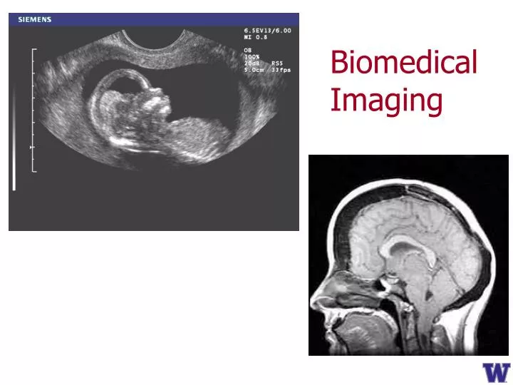
Biomedical Imaging
Molecular Imaging - Biomarker Localization Molecular Contrast Agent Image Beacon Target Protein Biological Targeting Molecular Image showing concentration of target protein
Why Ultrasound for Molecular Imaging? PET/CT MRI Ultrasound • Low cost • No radiation • Non-invasive • Highly portable • Real time imaging
Microbubble (1 mm diameter) Link Over-expressed specific cellular receptor Antibody to specific cellular receptor Targeted Ultrasound Contrast Agent
Light & Sound at Molecular Scale • Molecular imaging combining ultrasonics with photoacoustics
0 100 ns Photoacoustic Imaging Optical contrast at ultrasound resolution Reasonably large penetration depth (one way) 50um black bead Pulsed Laser Ultrasound detector
In-vivo PA imaging Photoacoustic image superimposed on ultrasonic image
Link Over-expressed specific cellular receptor Wavelength-dependent Optical absorber Antibody to specific cellular receptor Targeted Photoacoustic Contrast Agent
Targeted Photoacoustic Contrast Agent Endocytosis
Au Nanorods - Absorption Selectivity Prahl S, Oregon Medical Laser Center
IVC Aorta Right Renal Vein Left Renal Vein Surfactant (CTAB) Vascular Injury Cell Anti ICAM-1 ICAM-1 PAA ICAM-1 overexpressed Cell Bioconjugated Gold Nanorods Ultrasound-Photoacoustic Molecular Image Molecular PA image overlaid on top of US Image clearly showing site of vascular inflammation Potential Impact: US-PA imaging can guide over 1.5 M vascular interventions annually
US/PA Molecular Imaging Before injection After injection Pai-Chi Li - NTU
Molecular Therapy PA Temp – In vivo 20 x 10.5 mm 25 oC Temperature change 1 oC Stas Emelianov – UT-Austin
Are these signals nanoparticles or background ? PA Molecular Imaging: A Problem Tumor Before injection After injection
PA Imaging: A Problem Fundamental detection limit Tissue background PA Imaging: A Problem Saturation PA Signal Number of Particles / Voxel
Coupled Particles Coupled States Coupled States Coupled Agents: A Solution ?
Coupled States • Absorption modulation with light • Transient absorption vs. ground-state absorption • Differential-absorption photoacoustic (DAPA) imaging
PtOEP • Phosphorescence lifetime: hundreds of ns ~ tens of µs • Can be pumped at 532 nm • Can be used to estimate oxygen concentration with PA measurement
Imaging -43 dB
Transient Absorption vs. Pump Excitation probability
Excitation Probability vs. Depth (3 cm, 1− e−1) Oxyphor G2: σa,pmp = 1.9×10⁻¹⁶ cm2 at 633 nm
Conclusions: Coupled States • DAPA imaging can enhance PA contrast specificity by suppressing undesired background objects • DAPA imaging has the potential to provide sufficient imaging depths for biomedical applications
Coupled Particles Coupled Particles Coupled States Coupled Agents: A Solution ?
Background Tissue MNPs Coupled Magnetic Nanoparticles
Background Tissue MNPs Magnetic Field Coupled Magnetic Nanoparticles
Background Tissue MNPs Coupled Magnetic Nanoparticles
Differential PA Imaging • Image object with magnetic field off • Image object with magnetic field on • Use “motion” processing to differentiate regions of the image moving coherently with magnetic field
Differential PA Imaging Motion filtering mmPA imaging PA imaging Signal from coupled agent Signal from coupled agent Localized background Diffuse background
MNP-AU Core-Shell Agents Biomimetic synthesis of MNP-Au core-shell NPs using polypeptide as a template for directed Au nucleation and growth (Xiaohu Gao – BioE)
MNP-AU Core-Shell Agents • Core with finite magnetization • Shell with tunable optical absorption
Proof of Principle Pulsed laser UT water Preamplifier PVA FPGA GMNPs MNPs Rods Digitizer Electromagnet XY stage Function generator Current amplifier
g a PA image sequence mmPA image Motion tracking b Pixel-wise displacement f Weighting image c d Max. V+ e Max. V- Displacement fitting
Motion-enhanced PA Imaging Greater than 40 dB suppression
Conclusions: Coupled Particles • Developed core-shell nanoparticle with magnetic core and gold shell exhibiting high absorption in the NIR (Gao) • Nanoparticles can be manipulated with external magnetic field • Motion processing can dramatically reduce background PA signal
Challenge for a Potential Application What if the initial concentration of a targeted cell type circulating in the vasculature is too low to be detected and the PA background signal from blood overwhelms the PA signal from the targeted cells?
Fibre Laser pulses Magnet Photoacoustic signals Skin Transducer Blood vessel Acoustic waves Captured CTCs In vivo magnetic enrichment and multiplex photoacoustic detection of circulating tumour cells Ekaterina I. Galanzha1,2, Evgeny V. Shashkov1,3, Thomas Kelly1,4, Jin-Woo Kim5, Lily Yang6 and Vladimir P. Zharov1* 1Phillips Classic Laser and Nanomedicine Laboratories, Winthrop P. Rockefeller Cancer Institute, University of Arkansas for Medical Sciences, Little Rock, Arkansas 72205, USA 2Saratov State University, Institute of Optics and Biophotonics, Saratov 410012, Russia 3Prokhorov General Physics Institute, Moscow 119991, Russia 4Department of Pathology, University of Arkansas for Medical Sciences, Little Rock, Arkansas 72205, USA 5Department of Biological and Agricultural Engineering and Institute for Nanoscale Materials Science and Engineering, University of Arkansas, Fayetteville, Arkansas 72701, USA 6Winship Cancer Institute, Emory University School of Medicine, Atlanta, Georgia 30322, USA *e-mail: zharovvladimirp@uams.edu Nature Nanotechnology, vol. 4, pp 855- 860 (2009)
Contrast-enhanced Photoacoustic Imaging mm mm Congxian Jia1, Jinjun Xia1, Ivan M Pelivanov1,2, Sheng-Wen Huang1, Yongdong Jin1, Chi Hyung Seo1, Lingyun Huang1, Janet F Eary1, Xiaohu Gao1, and Matthew O’Donnell1 1University of Washington, USA 2Moscow State University, Russian Federation
Experimental Setup Pulsed laser Probe water + milk VeraSonics PVA MNP Dual Magnets tube XY stage Computer FPGA
Experimental Setup Pulsed laser Probe water + milk VeraSonics PVA MNP Dual Magnets tube XY stage Computer FPGA
Experimental Setup Pulsed laser Probe water + milk VeraSonics PVA MNP Dual Magnets tube XY stage Computer FPGA