Download
1 / 27
380 likes | 830 Vues
Biochemistry of cellular organelles. Kontinkangas, L101A. Lectures : 1. Membrane channels; 2. Membrane transporters ; 3. Soluble lipid/metabolite-transfer proteins ; 4. Mitochondria as cellular organelles ;

E N D
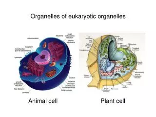
Biochemistry of cellular organelles Kontinkangas, L101A Lectures: 1. Membrane channels; 2. Membranetransporters; 3. Solublelipid/metabolite-transferproteins; 4. Mitochondria as cellularorganelles; Seminar: Isolation of subcellularorganelles; 5. Mitochondrialinheritance; 6. Mitochondria in health and disease; 7. EndoplasmicReticulum (ER) and lipids; 8. Structure and function of peroxisomes; Seminar: Mitochondria in cellular life. Dr. Vasily Antonenkov, Visiting professor Dept. Biochemistry, Oulu University Oulu, Finland Web site:
Lecture 7: Endoplasmic Reticulum (ER) and lipids Lecture content: • Morphology of ER; • Overview of functions; • ER and protein synthesis; • ER and lipid synthesis; • Lipid secretion out of cells; • Dolicholand protein glycosylation; • ER stress.
Morphology of ER • The most abundant membrane network in the cell, highly dinamic structure; • Rough (containing ribosomes) and smooth ER are membrane cisterna, vesicles and tubules held together by cytosceleton; • The cisternal space (or lumen, about 10% of the total cell volume) is continuous with the periniclear space but separate from the cytosol; • Smooth ER contacts physically with mitochondria and plasma membrane.
Protein sorting • Proteins synthesized on free ribosomes either remain in the cytosol or are transported to organelles; • Proteins synthesized on membrane-bound ribosomes are translocated into the ER when translation is in progress. They may be either retained within the ER or transported to peroxisomal membrane or the Goldgi and, from the the Goldgi apparatus, to lysosomes, the plasma membrane, or the cell exterior via secretory vesicles. • Proteins destined to ER contain a signal sequence at their amino (N) terminus; • Most signal sequences contain a stretch of hydrophobic aa, preceded by basic residues. The N terminus of growth hormone
Cotranslational targeting of proteins to ER • As the signal sequence emerges from the free ribosome, it is recognized and bound by the signal recognition particle (SRP, heterooligomeric protein); • The SRP escorts the complex to the ER membrane where it binds to the SRP receptor; • The SRP is released, the ribosome binds to a membrane translocation complex (Sec6) containing the channel protein that accomodates the signal sequence; • Translation resumes,and the growing chain is translocated across the membrane; • Cleavage of the signal sequence by signal peptidase releases the polypeptide into the lumen of the ER.
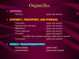
Rough ER function • The main function is the production and processing of proteins that are later exported from the cell or participate in formation of plasma membrane, lysosomes or peroxisomes; • Two types of newly synthetized proteins: (1) embedded into the membrane of ER, (2) soluble in the lumen; • Folding and modification of proteins using chaperon proteins in the lumen, initial glycosylation; • Transport of newly synthetized proteins mainly to the Golgi apparatus for further processing and export out of the cell; • Protein quality check - misfolded proteins sent back for recycling.
Smooth ER functions • Common: • Lipid (including steroids) synthesis; • Detoxificatrion – inactivation of harmful toxins, like drugs and metabolic waste; • Ca storage; • Transport of the newly synthesized proteins from rough ER by means of membrane vesicles. • Specific: • Glucose-6-phosphatase – gluconeogenesis; • Reproductive organs – production of steroid hormones.
Lipid synthesis in smooth ER Triacylglycerols • Fatty acid elongation pathway produces very long chain fatty acids from palmitic acid
Synthesis of membrane lipids: phosphatidylcholine as an example GPAT: Glycerol phosphate acyltransferase; LPAAT: Lysophosphatydic acid acyltransferase;CDP: cytidine diphosphate.
Lipid transfer : -through contact sites; -membrane receptors; -binding/transport proteins; -membrane transporters (ABC-proteins, others); -vesicular transport; -flipping. 10
Lipid traffic at membrane contact sites • (a) The ER forms a network for transfer lipids between organelles by using membrane contact sites (MCS), in these sites the lipid-transfer proteins (LTP) are active; • (b) LTP is shuttling between membrane receptors in the MCS and transfer lipid according to concentration gradient; • (c) The LTP may be simultaneously bound to both receptors of the opposite membranes of MCS that would increase the efficiency of lipid transfer. • MCS are also important to transfer Ca between organells.
Transfer of membrane lipids from ER to other organelles and to plasma membrane Two main mechanisms: The vesicles contain all components characteristic for membranes, including phospholipids, cholesterol and other lipids as well as some membrane proteins. In addition they can carry some proteins and/or metabolites confined inside the particles (e.g., excretion of proteins from epithelial cells during milk production in mammary glands). Soluble lipid-transfer proteins specific for membrane lipids such as phosphatidylcholine transfer protein.
Glycosylation • Glycosylation is the reaction in which a carbohydrate (glycosyl donor) is attached to a hydroxyl or other functional group of another molecule (lipids or proteins); • Protein glycosylation is a form of co-translational or post-translational modification; it is important in function of plasma membrane, cell to cell connection, activity of secreted proteins; • N-glycosylation – carbohydrate attached to a nitrogen of asparagine or arginine side-chains of proteins; it requires participation of a specific lipid – dolichol phosphate; mainly occurs in the rough ER; • O-glycosylation – carbohydrate attached to the hydroxy oxygen of serine, threonine, tyrosine, hydroxyproline, or to oxygens on lipids such as ceramide; mainly occurs in the Golgy apparatus; • Phospho-glycosylation – carbohydrates linked through the phosphate to a phosphoserine.
Dolichyl-phosphate is a glycosyl carrier lipid for N-glycosylation; • Dolichol is long enough to span the membrane 4-5 times; it is localized on the rough ER; • The oligosaccharide precursor is common to most eukaryotes, but after transfer to protein can be modified. Dolichyl-phosphate is formed by dolichol kinase: CTP + dolichol > CDP + dolichyl phosphate
Formation of lipid-linked oligosaccharide (llo) precursor The synthesis starts at the cytosolic face of the ER. (1-3) 2Acgl and 5 mannose residues are added one at time to DolPP; (4) Than the product is flipped to the luminal face; (5,6) In the latter reactions each mannose or glucose residue is transferred from nucleotide sugar to dolichol on the cytosolic face of the ER, then it is flipped and transferred to the growing oligosaccharide. The carrier (DolP) is then flipped back again to the cytosolic face (catalyzed by flippases).
Transfer from dolichol to protein • The oligosaccharide precursor is transferred from the dolichol to an Asn residue of the nascent protein, i.e. cotranslationally; • The Asn residue must be in a sequence N-X-S/T; • The process is catalyzed by oligosaccharide protein transferase. The enzyme is build up of three subunits. Two of them are ER transmembrane proteins. Their cytosolic parts bind to the larger subunit of the ribosome (they act as an anchor) The third subunit has the catalytic activity.
Modifications of N-linked oligosaccharides • After an oligosaccharide chain has been added to the protein, the 3 glucose and 1 mannose residues are removed by three different enzymes in a fixed order; • It occurs in the ER and is a signal that the protein is folded and can be transported to the Golgi for further processing. • The proper folding of the protein is controlled by the enzyme: UDP-glucose/glycoprotein glucosyl transferase (UGGT). If protein is misfolded, the UGGT reglucosylates it (adds glucose) that prevents protein from transfer to the Golgi.
Protein sorting in ER • Addition of glucose to proteins by UGGT trigger interaction of them with lectins – group of proteins able to interact with protein-bound sugars, such as calreticulin (CRT) and calnexin (CNX); • Interaction with CRT or CNX prevents from transfer of misfolded proteins from ER to the Golgi; • Delay in the transfer to the Golgi of misfolded proteins increases the probability of their recognition by glucose regulated protein 78 (Grp78) or EDEM protein that lead to excretion of misfolded proteins from ER. 18
Endoplasmic reticulum-associated degradation (ERAD) • The misfolded proteins are prone to aggregate and hence course an ER stress; • Some membrane-bound chaperones (i.e. EDEM protein) guide the retrotranslocation of the misfolded protein back into the cytosol; • In the cytosol the misfolded protein is ubiqutinated and subjected to proteasomal degradation; • The nature of the membrane machinery catalyzing export of misfolded proteins is not clear.
ER stress The consequence of a mismatch between the load of unfolded and misfolded proteins in the ER and the capacity of cellular machinery to cope with that. • The ER stress may results from deficiency of cell metabolism, i.e. deficit in energy supply, oxidative stress, etc.; • The ER stress leads to Unfolded Protein Responce (UPR). 20 20
Unfolded protein response (UPR) • UPR is an adaptive regulatory response of the cell to ER stress; • 3 mechanisms in mammals: ATF6, PERK, and IRE1; • All mechanisms eventually affect transcriptional (translational) regulation of proteins synthesis; • Results: • Increase in ER protein folding capacity; • Decrease in ER-dependent protein synthesis and folding.
IRE1 is a threonine/serine kinase and promotes cytoplasmic splicing of XBP1 mRNA, XBP1 is a transcriptional factor; • PERK is a protein kinase and active in attenuation of protein synthesis; • ATF6 is a transcription factor which undergoes maturation in ER; • All receptors somehow sense misfolded proteins in the ER lumen and transduce the signal to cytosol.
ATF6-dependent pathway • ATF6 is a transcriptional factor that is initially synthesized as an ER-resident transmembrane protein; • Upon accumulation of misfolded proteins, it is packaged into transport vesicles that deliver it from ER to the Golgi; • In the Golgi the ATF-6 protein is cleaved by two specific proteases leading to liberation of the N-terminal cytosolic domain ATF6(N); • ATF6(N) moves into nucleus to activate UPR target genes.
PERK pathway • PERK is an ER-resident transmembrane kinase; • When activated upon unfolded proteins, PERK oligomerizes and phosphorylate itself and translation initiation factor elF2 that inactivates it; • Hence, translation of most proteins decreases that helps cope with the influx of proteins in ER; • However, some proteins, including transcription factor ATF4, are preferably translated when elF6 is limiting; • ATF4 induces transcription of UPR target genes.
IRE1 pathway • IRE1 is a bi-functional enzyme (kinase/RNase) in the membrane of ER; • Due to binding the unfolded proteins, the IRE1 is oligomerized that activates kinase and endoribonuclease (RNase) activity; • RNase is active towards mRNA coding for transcription factor XBP1. It cleaves out one intron of this mRNA and after ligation (by other enzymes) this RNA translate XBP1; • XBP1 moves into nucleus to activate UPR target genes.
Suggested questions • Morphology of ER; • Sorting of the newly synthesized proteins and targeting of then into ER; • Functions of rough and smooth ER; • What kind of lipids are synthesized in ER and how (example); • What mechanisms of the lipid traffic do you know? Transfer of lipids at membrane contact sites; • Transfer of lipid from ER to other organelles and to plasma membrane; • N- and O-glycosylation of proteins in ER, role of dolichol; • Step-by-step mechanism of N-glycosylation; • Sorting folded from unfolded proteins in ER; • ER stress – what is it? Unfolded protein response – its functional role; • Three main mechanisms of the unfolded protein response –describe shortly them.
Thank you! Questions.