Erythrocytes aka RBC’s
620 likes | 1.89k Vues
Erythrocytes aka RBC’s. Laboratory Procedures. What is Blood?. Whole Blood = fluid and cells Fluid Component = PLASMA Cellular Component Erythrocytes (RBC) Leukocytes (WBC) Thrombocytes (Platelets). How Are These Cells Produced?.

Erythrocytes aka RBC’s
E N D
Presentation Transcript
Erythrocytes aka RBC’s Laboratory Procedures
What is Blood? Whole Blood = fluid and cells FluidComponent = PLASMA Cellular Component • Erythrocytes (RBC) • Leukocytes (WBC) • Thrombocytes (Platelets)
How Are These Cells Produced? Hematopoesis : the production of blood cells and platelets. Erythropoesis : the production of erythrocytes (RBC) In juveniles, blood produced in liver, spleen, thymus and bone marrow. In adults, primary site of erythropoesis is in the bone marrow. During times of hematopoetic stress, the slpeen and liver may produce RBC
Leukopoiesis : the production of leukocytes or WBC Thrombopoiesis : the production of thrombocytes or platelets
All of these cells come from ONE CELL!! STEM CELL Proteins called Cytokines are responsible in determining the fate of all stem cells.
These cytokines determine in the cell will be either : Myeloid : Erythroblasts – Erythrocytes (Erythropoietin) Megakaryocytes –Platelets Myeloblasts –Leukocytes / Monocytes
Blood Composition Separates into three components: Red Blood Cells (RBC’s) White Blood Cells and platelets (buffy coat) Plasma Bottom 1/3 to ½ of tube contains the heaviest of cellular material (the RBC’s).
Hematocrit=PCV (Packed Cell Volume) To determine hematocrit, whole blood is centrifuged to pellet the red blood cells. Plasma remains on the top of the red cells. The fraction of blood that is packed is the hematocrit and is read as a percentage.
Hemoglobin (Hgb) Normal values are usually 1/3 of the hematocrit. Each hemoglobin molecule has 4 heme units attached to globulins. Abnormal heme groups, cannot carry oxygen. Carboxyhemaglobin- Hgb has a higher affinity for CO than O2. Bright red blood Methemoglobin- The Fe molecule is oxidized to Fe+3. Blood becomes brown. Tylenol toxicity in cats.
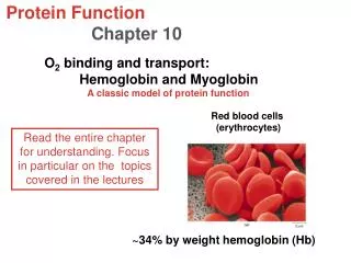
Red Blood Cells Function: Carry oxygen to the tissues Oxygen must be carried at enough pressure to permit rapid diffusion of oxygen. The RBC is a vehicle for hemoglobin which is the carrier molecule for oxygen. The sigmoid shape of curve is a result of the cooperative binding from the 4 hemoglobin molecules.
Erythrocytes The morphological features of mature red blood cells of dogs, cats, horses, and ruminants are generally very similar in that they all lack nuclei, stain reddish to reddish-orange. Erythrocytes are biconcave discoid-shaped cells.
The major differences are in the size of the red blood cells and the degree of central pallor. Listed from largest to smallest in size are : Dog Cat Horse Cow Sheep Goat
Erythrocytes Maturation of a RBC. Only occurs in the bone marrow of normal adult animals. Occurs in the spleen and liver of the fetus. Maturation time usually takes 5 days. Regulated by erythropoietin (EPO) which is increased in the presence of hypoxia. In most species, the kidney is the sensor organ and major site of EPO.
Red Blood Cells (Erythrocytes) No nucleus due to have to fold and squeeze through tight spaces. Normocytes- cells look normal
Erythrocyte Life Span Dog- 110 days Cat- 70 days Cow- 160 days Horse- 145 days Man- 120 days Mouse- 30 days
Erythrocyte Life Span Stem Cell → Rubriblast → Prorubricyte → Metarubricyte → Rubricyte → Reticulocyte → RBC Rubricyte- nucleated RBC releases in severe anemia. No more mitotic division takes place after this stage. .
Normal Erythrocytes • Morphologic features vary from species to species • Dogs: Biconcave disk shape with central pallor • Cats: Generally round with little central pallor. • Birds: Contain a nucleated RBC
Categories of Erythrocyte Characteristics 1. Cell arrangement on the blood film. 2. Size 3. Color 4. Shape. 5. Presence of structures on erythrocytes
Cell Arrangement on Blood Film • Rouleaux • Rouleaux formation is a group of erythrocytes in stacks. This can be a sign of increased fibrinogen or globulin concentration secondary to inflammation. • It can also be an artifact seen in blood that is held too long before preparing the blood slide or in blood that has been refrigerated. Can be a normal occurence!! Most Commonly Seen in Horses!!!
Cell Arrangement Continued • Agglutination • Agglutination, which appears as rouleaux, occurs in immune-mediated disorders. An antibody coats the cell causing bridging or clumping. • If you add a drop of saline to a drop of blood rouleaux formation will disperse and agglutination will not
Size • Terms: • Anisocytosis: • Variations in size • Can indicate anemia • Macrocytosis: • Larger than normal cell size • Liver disease or Vitamin B12 deficiency • Microcytosis: • Smaller than normal cell size • Iron deficiency
MCV • Mean Corpuscular Volume • Describes cells as normocytic, microcytic, or macrocytic. Calculates the average volume of rbc’s. • MCV=(Hematocrit x 10)/RBC count in millions • Normal: 66-77
Color • Polychromasia (polychromatophilic): • Polychromatic erythrocytes exhibit a bluish tint. The tint is due to a small amount nucleus retained in the cytoplasm. These are young cells and may appear as a reticulocyte • Hypochromasia: • is a decrease in color, due to a decreased staining intensity caused by insufficient hemoglobin within the cell. • Iron deficiency is the most common cause. • Hyperchromasia (hyperchromatophilic): • refers to cell that appears darker than normal cells. This gives the appearance that the cell is over saturated with hemoglobin. The erythrocyte has a fixed maximum capacity for hemoglobin and over saturation can NOT occur.
MCHC • Mean Corpuscular Hemoglobin Concentration • describes cells as normochromatic or hypochromatic. • MCHC= (Hgb)/(Hct) x 100 • Normal is 31-36% • (we will come back to this calculation again)
Hypochromasia Usually associated with iron deficiency. Especially in Llamas
Hypochromasia continued • Hypochromatic should be differentiated from cells with the center “punched out”. A punched out appearance can be an artifact due to improper smear technique
Shape • Poikilocytosis • Poikilocytosis is a major deviation in the normal shape of the erythrocyte. The term poikilocytosis is an umbrella term that is used for any and all abnormally shaped erythrocytes and does not suggest a specific diagnosis
Schistocytes (Fragmented Cells) Also known as poikilocytes. RBC’s with abnormal shape. Formed as a result of shearing of the cell by fibrin strands. This occurs when red blood cells rapidly pass through microvasculature that is lined or meshed with strands. They are observed in fragmentation hemolysis caused by DIC, vascular neoplasia, endocarditis, and possibly iron deficiency anemia.
Acanthocytes (Spur Cells) • The term acanthocyte is derived from the Greek word “acanthi” meaning “thorn” Acanthocytes are cells with five to ten irregular, blunt, finger-like projections. • The projections with vary in width, length and surface distribution. These cells are seen in animals with altered lipid metabolism such as cats with hepatic lipidosis or dogs with liver disease.
Echinoctyes (Burr Cell) • Echinocytes have multiple, small, delicate regular shaped spines evenly distributed around the cell and are indistinguishable from artificially crenated cells.
Echinoctyes Continued • Echinocyte formation can be artificial, often seen with slow drying blood films or if the EDTA tube was underfilled. This artifact is then termed crenation. • Echinocytes have been associated with renal disease, lymphosarcoma and rattlesnake bites in dogs. • They can be seen after exercise in horses.
Crenation Identified as the presence of many irregular membrane projections involving most RBC’s. It is usually an artifact due to slow drying of the blood film. Commonly observed in pig blood but can be seen in any species.
Drepanocytes (Sickle cell) • These cells are crescent shaped with pointed ends. • Drepanocytes are often seen in normal blood of deer and goats. It is thought to be a result of low oxygen tension.
Keratocyte (Helmet/Blister Cells) • Also called blister cells or bite cells. Keratocytes are associated with trauma especially cellular damage from contact with fibrin strands.
Prekeratocytes • Cells with pseudovacuoles are called blister cells or pre-keratocytes.
Spherocytes Cells have a spheroid shape instead of the usual biconcave disk shape. Have reduced cell membrane and are hypochromatic. Seen most frequently in autoimmune hemolytic anemia (AIHA). When WBC partially remove antibody-coated membranes. Usually seen in dogs.
Stomatocytes • The appearance of stomatocytes with their oval or rectangular central pallor has been compared to a smiling face, a fish mouth, and a coin slot. • Stomatocytes are associated with an hereditary condition but are also seen in liver disease, acute alcoholism (humans), and electrolyte imbalances.
Anulocytes • These are bowl shaped erythrocytes that form as a loss of membrane flexibility that does not allow the cell to return to a normal shape after passing through a capillary. They can occur due to lowed hemoglobin concentration or as an artifact.
Dacryocytes (tear drop cells) • These tear drop shaped cells are seen in myeloproliferative diseases. These cells can be produced as an artifact but can be identified by the direction of their tail
Dacryocytes produced as an artifact have their tails pointing in the same direction.
Target Cells and Folded Cellsaka Codocytes Two types of cells observed mainly in dogs. Represent cells with an outfolding of the rbc membrane. The cell membrane is thin and flimsy. Can be associated with liver dz and reticulocytosis.